Podcast 77: Gait analysis, Forefoot Running & more.

Plus, the 5 neurologic gait compensation expressions.

*Show sponsor: www.newbalancechicago.com

A. Link to our server: 

http://traffic.libsyn.com/thegaitguys/pod_77final.mp3

Direct Download: 

http://thegaitguys.libsyn.com/podcast-77

B. iTunes link:

https://itunes.apple.com/us/podcast/the-gait-guys-podcast/id559864138

C. Gait Guys online /download store (National Shoe Fit Certification and more !) :

http://store.payloadz.com/results/results.aspx?m=80204

D. other web based Gait Guys lectures:

www.onlinece.com   type in Dr. Waerlop or Dr. Allen,  ”Biomechanics”

______________

Today’s Show notes:

Google X acquires ‘tremor-canceling spoon’ startup
http://venturebeat.com/2014/09/10/google-x-acquires-tremor-canceling-spoon-startup/

The 5 expressions of neurologic gait decomposition,
Last week we did an online teleseminar … . .
An acoustic startle alters knee joint stiffness and neuromuscular control
http://onlinelibrary.wiley.com/doi/10.1111/sms.12315/abstract
Effectiveness of Off-the-Shelf, Extra-Depth Footwear in Reducing Foot Pain in Older People: A Randomized Controlled Trial
http://biomedgerontology.oxfordjournals.org/content/early/2014/09/08/gerona.glu169.abstract
reader:
I really appreciate learning from you!! I have a bit of a loaded question that I will try to explain clearly to the best of my ability. About 2 years ago, I broke my left shin (hairline-fibula) in a MMA fight. After it healed, a few things have been happening that I assume are connected but can’t quite put my finger on. My ankle mobility on my left ankle is worse than my left. I seem to have permanent turf toe as well. My right glute, ham, and erector are hyperactive.
Additionally, many times when sprinting, pushing a sled, etc, my right quad will become fatigued much more than my left. I believe it’s because I’m not fully extending my left ankle, and relying on my right leg more. Whenever I squat or deadlift, I feel similar too. The right glute and erectors get much more of a “pump” than my left. With all of this, is there anything you would recommend? I truly appreciate it!! It is very frustrating. Thank you again!


People tend to forget about the peroneal muscles. This is what it looks like when the brain forgets.

This client came to see us for obvious reasons but the case details are not what we are focusing on today. Gait gets pretty messed up when a critical component or phase is lost or forgotten.  

In last weeks teleseminar on www.onlineCE.com we discussed several gait cases. In these cases 5 things kept coming up when it came to looking at (specifically) neurologic gait compensations:

  1. slowing of gait
  2. wider based gait
  3. increased ancillary movements 
  4. utilizing support when needed or available
  5. shorted step length and stride length

In this video, it is clear that this person has some serious neurologic problems engaging the peroneal muscles and controlling ankle and foot function and as a consequence you see evidence of some of the itemized issues above, namely, calculated movements, nearly zero arm swing and step length from left to right is abbreviated. 

It can go both ways. The neurologic problem can affect one’s gait, but one’s resultant gait can then affect cortical function, driving an endless loop. Recently, five studies presented at the Alzheimer’s Association International Conference in Vancouver Canada provided striking evidence that when a person’s walk gets slower or becomes more variable or less controlled, his cognitive function is also suffering.(2)  

A person’s gait and their neurologic function cannot be separated. The stuff just run’s too deep.  This is why we love gait so much, because to fully understanding someone’s clinical problems we must understand how and why they move.  There are clues in everyone’s gait that can help you clinically. The question is, will you notice them ? Do you know what normal gait is to begin with ? Will you understand what you are seeing and realize it is a compensation? Will you fix what you see or look deeper to find the cause of what you see ? 

Shawn and Ivo,

The Gait Guys

Gait Posture. 2013 Jul;38(3):549-51. doi: 10.1016/j.gaitpost.2013.02.008. Epub 2013 Mar 11.

Altered gait termination strategies following a concussion.

Buckley TA1, Munkasy BATapia-Lovler TGWikstrom EA.

2.  http://www.nytimes.com/2012/07/17/health/research/signs-of-cognitive-decline-and-alzheimers-are-seen-in-gait.html?_r=1&

This is part 2 of a 2 part post; with the video from the case previously discussed

please note the following in the video:

  • body lean to left during left stance phase (to clear right longer leg)
  • circumduction of right lower extremity  (to clear right longer leg)
  • lack of arm swing bilaterally (cortical involvement)
  • patient looking down while walking (decomposition of gait)
  • shortened step length (decomposition of gait)
  • increased tibial varum bilaterally

ASSESSMENT:  This patient’s short leg and internal tibial torsion impediments to her full recovery. She has increased tibial varum noted which is complicating the picture. This is causing pathomechanics and an abductory moment not only at the knee but also in the lumbar, thoracic and cervical spines.

WHAT DID WE DO?:                    

  • We attempted to do the one leg standing exercise. She needed to hold on and did not feel stable on the left hip while performing this.  This is probably more of confidence rather than ability issue. 
  • We gave her the stand/sit exercise to try to improve gluteal recruitment.
  • We also gave her the lift/spread/reach exercise to attempt to strengthen her feet.
  • A full-length 5 mm lift was cut for the left shoe  She felt more stable when walking on this.
  • She was treated with IC, PIR and manipulative therapy and neuromuscular stim of the knee as well as left hip area above, below and at the joint line of the knee as well as gluteus medius and minimus.   
  • We may need to consider building a more aggressive orthotic with a forefoot varus post depending upon her progress and response to care  

 The Gait Guys. Making it real, each and every post here on the blog.

special thanks to SZ for allowing us to publish her case, so others can learn

tumblr_nc9grebaIQ1qhko2so1_1280.jpg
tumblr_nc9grebaIQ1qhko2so2_1280.jpg
tumblr_nc9grebaIQ1qhko2so3_1280.jpg

Wow! What would you do?

This is part 1 of a 2 part post. Look for the other one a few minutes after this one with a video up top for the conclusion

PRESENTING PROBLEM: This 54 YO female patient presents with with left sided knee pain.  She had a total knee replacement (TKR) done in 2011.  She’s had a significant amount of discomfort on the medial aspect of the knee since then. She had an MRI of the hip done thinking the problem was there, and found nothing.   She is walking with a bad limp, left leg is half inch shorter than the right.  Pain is worse at night, changes with weather. 

She has knee pain on the lateral aspect (points to tibial plateau and joint line) with swelling that goes down to the ankle left side.  She has been wearing a “Good Feet” OTC orthotic on the left side which she states helps quite a bit.

Generally speaking, stretching and analgesics make the discomfort better.    Ibuprofen 400 mg. b.i.d. can take the edge off  Soft sided brace (neoprene sleeve) makes a difference as well. The hard sided brace gives her difficulty.

WORK HISTORY: She works for a preschool.  Her job involves standing and getting up and down a lot.  

FAMILY HISTORY:  She has left sided lid ptosis, this evidently is familial.  

PHYSICAL EXAM:  She stood 5’ 1” and weighed approx. 150 pounds.

Viewing the knees bi-lat., the left knee is markedly externally rotated.

She does have a left short leg; tibial and femoral.  She has bilateral tibial torsion (look at the tibial tuberosities and drop a line straight down; it should pass through the 2nd metatarsal head) and marked internal tibial torsion on the left side (>60 degrees) with femoral retrotorsion (less than 8 degree angle of femoral head with the shaft) on this side.  There is no rotation of the thigh or leg past zero degrees midline. .  She had 10 degrees of tibial varum on the left hand side.  Her Q-angle is 10 degrees on that side.  There is plantar flexion inversion of the foot.  Left lower extremity has less sensation secondary to the her TKR  surgery.

Gait evaluation reveals a fair amount of midfoot pronation noted on the left hand side in addition to an intoed gait.  She has to lean her body over to the left to get the right leg to clear.

Some mild weakness noted of hip abduction musculature left hand side gluteus medius, middle and anterior fibers. Knee stability tests were negative.

Neurologically, otherwise, she had full integrity with respect to sensation, motor strength and deep tendon reflexes in the upper and lower extremities.

Please see part 2 of this post for additional info including our assessment and what WE did.

 The Gait Guys. Making it real, each and every post here on the blog.

special thanks to SZ for allowing us to publish her case, so others can learn

Podcast 76: The FMS™ screen and Injuries, Impact Loading & more.

Podcast 76: Association of Functional Movement Screen™ With Injuries, Wool workout gear, landing softly and more !

*Show sponsor: www.newbalancechicago.com

A. Link to our server: 

http://traffic.libsyn.com/thegaitguys/pod_77.1_76final.mp3

Direct Download: 

http://thegaitguys.libsyn.com/podcast-76

B. iTunes link:

https://itunes.apple.com/us/podcast/the-gait-guys-podcast/id559864138

C. Gait Guys online /download store (National Shoe Fit Certification and more !) :

http://store.payloadz.com/results/results.aspx?m=80204

D. other web based Gait Guys lectures:

www.onlinece.com   type in Dr. Waerlop or Dr. Allen,  ”Biomechanics”

______________

Today’s Show notes:

 
Last week on our social media sites we posted this article that garnered 9000+ hits:
Runner? LONG DISTANCE runner? Better be careful out there!
http://www.sciencedaily.com/releases/2012/06/120604093108.htm
 
then this news this week:
Well-Regarded Endurance Athlete Chad Denning Dies While Running Appalachian Trail | Valley News
 
Association of Functional Movement Screen™ With Injuries in Division I Athletes
 
from a reader:
Hey guys, great site, sometimes a bit more than I know at this point. Just graduated from massage school in april. I have been diagnosed with tendonosis of the Achilles heel. Also finding that my leg doesn’t fully extend while walking, anything I can do besides hamstring and calf stretches. It really happened after a 30 mile hike with a 40 lb backpack, Help 
Thanks, sincerely Hector
Synthetic Workout Gear Smells Worse Than Cotton Gear
 
 Land Softly And Carry Less Injury Risk

http://running.competitor.com/2014/07/injury-prevention/land-softly-and-carry-less-injury-risk_11174


tumblr_nc1x994UP81qhko2so1_1280.png
tumblr_nc1x994UP81qhko2so2_1280.png

It is marathon season in Chicago. Time for this old reader-favorite blog post on Black toe nails !  

The Black Plague (ok, kinda sort of)……Subungal Hematomas in Runners. Blood under the toe nail.  It is not exactly what you think it is !

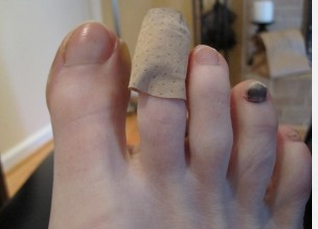
There are two pictures here, cursor to the right and see the slider that will toggle between the two photos. The photo with bandaid credit given towww.healthandrunning.com the other photo……is a runner client of ours with both a callus pattern on the tip of the 2nd toe and an early small subungal hematoma (read on !)

__________________________________________________________________________

We get inquiries about the black toe nail “Syndrome”……aka…..blood under the toe nails in our runners, and how to avoid them. Lets look at this phenomenon as it pertains to the foot.

This problem has a clinical name, “subungual hematoma”. It means a collection of blood under the finger or toe nail.  There are many causes of the subungal hematoma (SH for short as we move forward here).  Here are a few, but we have yet to find any good journal articles for one cause that we are seeing as a possible cause…one we will discuss here shortly. 

One cause is obvious, the crush injury where someone steps on your toe, you drop something onto it or smash it into something.  This is something we have all done at some time. 

 The most commonly theory of cause is repetitive trauma, thought to be that of repeated impact of the toe into the top or end of the shoe.  Heim et al  noted this in 2000.  This really got us to thinking.  Why, when we see these SH’s, do the runners never seem to have shoes that are too short / small or shoe signs of friction (wear patterning) of the toe nail into the top of the shoe’s upper ?  Often the runners insist there has been no such contact within the shoe.  So we started our own investigation making sure to ask all our runners what they thought and felt as they ramped up their miles in prep for marathons and 20 mile runs or daily doubles, particularly those who seemed regularly susceptible to SH’s.  We will discuss our findings and thoughts momentarily, but lets get back to some of the more well known information on SH’s.

The medical literature is full of other types of causes or clues of SH’s that must be investigated, such as medication reactions, autoimmune skin disorders, melanoma, blood disorders (dyscracias or clotting problems). These certainly are not the norm.

It is important to know the anatomy of the area because the nail bed is very rich in vasculature (hence the hematoma creation) and nerve endings (hence the pain) when blood collects in the confined area or it gets torn off from trauma.  The nail bed is a derivative of the epidermis containing keratin which gives it its hard nature. The nail grows from a nail root in front of the cuticle and grows distally at a slow but (usually) steady rate.  This area is frequently susceptible to fungal infections which destroy the tissue in the area and possibly make SH’s more common.

We will not get into the aggressive treatment of things here because that is 1) not our purpose here and 2) we do not want to be accountable for people getting infections  from boring a hole into the nail bed (trephine) to release the blood or the consequences of using plyers to yank it off.  We just tend to recommend they be left alone and let nature take its course.  (For those bold and tough gang, who chose the plyers method, you should know that there is no fatty tissue beneath the nail and the underlying bone to cushion the area, the nail is the only protection; furthermore you should know that the extensor tendon attachment is awfully close to the proximal nail bed root area !).  But when pain it too much, we have our people we refer these cases to.  Rather, we tend to look for a cause of the problem. 

In a limited number of cases we do see a shallow toe box where there is little room for toe extension, thus the nail can get rubbed on the roof of the toe box repeatedly causing a lifting action of the nail from its vascular bed.  This a more plausable cause in our opinion over the “toes hitting the end of the shoe” phenomenon put out there by many sources.  Particularly when most people size their shoes sufficiently long enough for the distal foot slip migration that occurs at mid-foot load within the shoe.  In  these cases a close cropping of the toe nail shoe stop the lifting/friction phenomena on the toe box roof. 

However, we seem to be seeing a more frequent trend that we wanted to share  here.  It seems to go hand in hand with the plague of flexor dominance in our society these days.  What we are seeing is a predominance of toe flexion (either a gentle or marked toe flexion ….we sometimes refer to it as toe hammering) in our runners.  This just makes sense because of the posterior compartment dominance in runners.  (The posterior compartment is made up of the gastroc-soleus complex, long toe flexors and tibialis posterior).  So if this compartment is dominant, and there is not sufficient home work to off set the flexor dominance with extensor exercises, then this flexion dominance will continue and possibly worsen.  As you will see either in yourself, our photo here, or on the feet of many of your co-runners is a distal “tip of the toe” callus development (usually most on the second toe, and less moving into the more lateral toes) immediately below the leading edge of the toe nail.  This callus coincides well with a distal gripping phenomenon of the long flexors (Flexor digitorum longus). So, now imagine, to get the callus there must be repeated friction and since the toe is not hitting the end of the shoe it must be friction into the sock liner bed of the shoe. And if this is the case, the skin is pulled at a differential rate over the distal phalange than the nail bed there will be a net lifting response of the nail from its bed as the skin is drawn forward of the backward drawn phalange  (put another way, the callused toe tip is fixed to the sock liner for grip, and then the phalange is drawn backwards from this contact point creating a NET movement of skin forward thus lifting the nail from its bedding).  [For an at-home example of this, put your hand flat on a table top. Now activate your distal long finger flexors so that only the tip of the fingers are in contact with the table top.  Now, without letting the finger tip-skin contact point move at all, go ahead and increase your long flexor tone/pull fairly aggressively. I defy you to not feel some  pressure building under the distal tip of the finger nail as the skin is RELATIVELY drawn forward.]   And with the nail bed being so vascular, micro bleeding can occur.  This bleeding is slow and takes time.  Which brings the big question to light, SH’s seem to mostly occur on very long runs, and never on short runs (where there is not enough nail bed separation repeatedly to create enough damage to bleed, not to mention fatigue of the other toe/foot intrinsic muscles thus necessitating more use of the more powerful long toe flexors.)

There  does not seem to be anything out there in the information on this supposition.  Maybe we are crazy…….but we do see alot of runners.  And once we bring the awareness of the problem to our runners and show them  how to reduce the flexion dominance with exercises to gain more extension balance, do we see an arrest of any further Subungal hematomas. 

We would love to hear your thoughts and experiences with them, both clinically and as a runner. Let us know what you think about our plausable cause.  

we remain……The Gait Guys

Remapping the Cortex: How Rehab Exercise does it.

Below are two studies that we recently incorporated into 2 neurologic gait cases during one of our global teleseminars on www.onlineCE.com.  You can find that lecture there in a few weeks but we have dozens of our other presentations available there presently. 

Injury to a body part starts a reorganization of the brain cortex. We know this occurs from a plethora of studies but most of them are based on injury induced changes and not from treatment-induced means.  These studies support the treatment induced changes that occur in the central nervous system, and they are profound and give us comfort and validity in our work. The findings of these studies should not be a shock to you if you are in the work of manual therapy and rehab. 

The one study used transcranial magnetic stimulation to map the cortical motor output area of a hand muscles on both sides in 13 stroke patients in the chronic stage of their illness before and after a 12-day-period of constraint-induced movement therapy.

What they found was “post treatment the muscle output area size in the affected hemisphere was significantly enlarged, corresponding to a greatly improved motor performance of the paretic limb”. As the study showed, this suggested a recruitment of adjacent brain areas. Even at 6 month follow up examinations “the motor performance remained at a high level, whereas the cortical area sizes in the 2 hemispheres became almost identical, representing a return of the balance of excitability between the 2 hemispheres toward a normal condition.”

The second study (2) looked at limb immobilization in 10 right-handed subjects with right upper extremity injury that required at least 14 days of limb immobilization. Subjects underwent 2 MRI examinations post injury, 48 hours and 16 days post immobilization. Cortical thickness of sensorimotor regions and FA of the corticospinal tracts was measured.  The findings showed “a decrease in cortical thickness in the left primary motor and somatosensory area as well as a decrease in FA in the left corticospinal tract. In addition, the motor skill of the left (noninjured) hand improved and is related to increased cortical thickness and FA in the right motor cortex.”

These studies suggest the findings are associated with skill transfer from the right to the left hand. It was suggested that immobilization induces rapid reorganization of the sensorimotor system. 

Rehab works, but everyone here on The Gait Guys already knew that. It is just nice to know the specifics of “how”.  

Please go to these articles and get the specifics for yourself. Don’t take our word for it ! 

references:

1. Stroke. 2000 Jun;31(6):1210-6.Treatment-induced cortical reorganization after stroke in humans. Liepert J1, Bauder H, Wolfgang HR, Miltner WH, Taub E, Weiller C.

2. Langer N, et al “Effects of limb immobilization on brain plasticity"Neurology 2012; 78: 182–188.


Steppage gait ? Or just a runway model ?  Take the thinking farther.
Today we have a short blog post for you. You may take the topic simply on the surface or cogitate over it and find some deeper epiphanies from the well of knowledge we have tried to present here on our blog for the past 4+ years.  
It is clear that in this video that the model has a consciously driven steppage gait. Meaning, she is lifting her limb/foot via exaggerated hip flexion and knee flexion to clear the foot.  This is often seen unilaterally in a foot drop case where the client has a neurologic lesion that for one reason or another has impaired the client’s ability to extend the toes or dorsiflex the ankle sufficiently to clear the foot (so they do not drag toes and trip/fall).  
But, why is she doing this steppage gait ? It is highly unlikely that she has bilateral lesions.  Sure, she was asked to walk this way by her mentor but again, take it further.  Is there a factor making this gait necessary regardless of the coaching ? 
Obviously the answer is yes or we wouldn’t be doing a blog post on this topic.  She is wearing ridiculously high heels. This is forcing her into an extreme plantarflexed foot and ankle posture. IF she were to swing her leg normally during the swing phase she would drive the foot and ankle into dorsiflexion (a normal gait event) and the long pointed heel would be made more prominent as it was driven forward and downward. This would surely catch on the ground, immediately driving the foot into sudden violent forefoot loading and pitch her into a forward fall.  Yes, you have seen this on the run way videos on youtube, and yes we know you laughed too ! You see, when wearing heels this high, one must deploy a certain degree of steppage gait to clear the heel because ankle plantarflexion is fraught with the risk we just discussed above, the heel is too prominent and will catch. How much steppage (knee flexion and hip flexion to clear the foot) is necessary ? Well, to a large degree it depends on how much of a heel is present.  If you are wearing a small heeled shoe, lets say 1 inch, then a small steppage is necessary.
None the less, there is a bigger problem lurking and brewing underneath when heels are a regular occurrence. Slowly and gradually the disuse of the anterior compartment muscles (Extensor dig., Ext. hallucis, peroneus tertius, tibialis anterior) will weaken and the posterior compartment will shorten respectively. IF left too long, it will result in tightness (yes, there is a difference between tightness and shortness, one is a neurlogical protective mechanism, the other is a more permanent change.) We have said this many times here and in our videos, much of posterior compartment problems (ie achilles tendonitis, Sever’s, Hagglunds etc) are related to a degree of anterior compartment weakness, skill deficits or endurance challenges.  Wearing high heels often will often, but not always, increase this risk. 
If you are an athlete, but someone who wears high heels often, you may have to do extra work to keep your anterior compartment competent on several levels.  Eccentric strength is just as important as concentric in this region. Remember, many gait problems come on slowly, a slow simmering smoldering fire. And remember this last point about heeled shoes, your forefoot is always being loaded initially in ankle plantarflexion, this is not normal and in time this will have a cost in many people.  
One last thing. We are not necessarily talking about dress shoes, although they are a greater culprit.  Many running shoes still have accentuated rear foot stack heights where the heel will be many millimeters above the plane of the forefoot.  Do not discount these shoes as a possible contributor of your problem, remember, physiological adaptation takes time to express into a biomechanical symptom creating problem, and it may take quite some time to resolve your compensations and adaptations.
PS: drive that “cross over gait” lady.  Fools.
Shawn and Ivo
the gait guys
Subtle clues often provide the answers.
We like yoga as much as anyone else. We saw this picture on the latest cover and couldn’t resist making a few comments on this pose.
Yoga has many benefits. Our understanding is that in addition to the c…

Subtle clues often provide the answers.

We like yoga as much as anyone else. We saw this picture on the latest cover and couldn’t resist making a few comments on this pose.

Yoga has many benefits. Our understanding is that in addition to the cognitive and spiritual effects of yoga, is that it helps to build your core.

 At first look you may say that this woman has a few issues:

  • she has a right pelvic shift and a left body lean
  • She has slight head rotation to the right and a slight left head tilt
  • you may have noticed that she appears to have more tone in the musculature on the right side of her face than on the left.   Just look at the nasolabial fold as well as the corner of her mouth any area of wrinkling underneath her left orbit.
  •  You may have also noticed the subtle flexion and lack of external rotation of the right hip.

 You may go on and think that she has a week right gluteus medius as well as an overactive quadratus lumborum on the left-hand side which may be causing the pelvic shift. The head tilt may be in compensation for the right side gluteus medius weakness and the subtle rotation may be an attempt to engage a tonic neck response. ( a tonic neck response is  ipsilateral extension of the upper and lower extremity to the side of head rotation with contralateral flexion of the same counterparts.

 You may have also noticed that the toes of the right foot are not dorsiflexed and that her hair appears to be flowing on the right side, and this is not the case at all, but rather she is either standing on a sloped surface or on the downward phase of a jump. According to the magazine it is the latter.  If you caught this at first then congratulations: you are sharper than most. If not remember to always look for subtle clues.

 Like Sir Topham Hat says in Thomas the Train: “  You didn’t get the whole story. What really happened is what really matters.

So why the mild facial ptosis on the left side? She could have had an old Bells palsy, or other form of facial paresis. Note that mostly the lower portions of the (left) face are affected (ie, below the eye). We remember that the upper portions of the face receive bilateral innervation but lower portions of the face unilateral innervation, from the contra lateral facial motor nucleus; this is why it could be a mild upper motor neuron lesion (micro infact, lack of cortical afferent input) and not an lower motor neuron lesion (like Bells Palsy). Why is this germane? Or is it not?

Stand in front of a mirror. Jump up in the air trying to assume the same pose as this woman does and what do you see.  Make sure that you jump up from both legs and then bring one leg over and your hands in front of you in the "praying position”. You may want to have a friend take a snapshot of you performing this. You will notice that you have contralateral head rotation,  a pelvic hike on the side opposite the leg that’s extended and a head tilt to the side that is flexed.  You are attempting to stabilize your core as you’re going up and coming down.

What we are witnessing is a normal neurological phenomena.  This gal merely seems to have some limited external rotation of her left hip. Now perform the same maneuver again but this time don’t externally rotate your leg as far as this woman does and what do you see. You should’ve seen an increase in the aforementioned body postures.

Subtle clues are often the key. Keep your eyes and ears open. 

The Gait Guys. Helping the subtle to become everyday for you, with each and every post.

Podcast 75: Joint Symmetry, Clinical Pearls & Random Thoughts

Lots of good random topics on today’s podcast, including possible causes of leg length discrepancies.

*Show sponsor: www.newbalancechicago.com

A. Link to our server: 

http://traffic.libsyn.com/thegaitguys/pod_true76f.mp3

Direct Download: 

http://thegaitguys.libsyn.com/podcast-75-joint-symmetry-cases-stuff

B. iTunes link:

https://itunes.apple.com/us/podcast/the-gait-guys-podcast/id559864138

C. Gait Guys online /download store (National Shoe Fit Certification and more !) :

http://store.payloadz.com/results/results.aspx?m=80204

D. other web based Gait Guys lectures:

www.onlinece.com   type in Dr. Waerlop or Dr. Allen,  ”Biomechanics”

______________

Today’s Show notes:


Neurons in human skin perform advanced calculations

http://medicalxpress.com/news/2014-09-neurons-human-skin-advanced.html

RunScribe Is A Wearable For Granular Gait Analysis
Free dialogue on leg length discrepancies.

Evidence for joint moment asymmetry in healthy populations during gait.

Gait Posture. 2014 Jul 1. pii: S0966-6362(14)00610-9. doi: 10.1016/j.gaitpost.2014.06.010. [Epub ahead of print]
The contralateral foot in children with unilateral clubfoot, is the unaffected side normal?

http://www.gaitposture.com/article/S0966-6362(14)00523-2/abstract

Gait guys case on Club foot:

http://thegaitguys.tumblr.com/post/23230149195/we-could-have-easily-made-this-a-blog-post-about

How injury and pain reorganize the brain.

Gait, Arm Swing and Reorganizing the Brain

When we injure a body part there is a price to pay, how expensive it will be is entirely up to you. Upon injury, the brain takes note and typically dives into a backup plan of neurologic inhibition, neuro-protective tightness and alteration of motor patterns to protect that injured area and allow it to heal. Moderating and altering the forces and demands upon said tissues is the goal to enable healing, if we as humans, don’t get in the way first (“I have to get that run in, I am behind in my training with all these injuries !” or “Ah, its still not that bad, the pain wasn’t worse on Tuesday’s run, I will be ok.”). The bigger question for most folks is, will you listen to what your body is asking of you? Heed the warnings and messages, and your injury will come and go in a timely manner, ignore the messages and welcome to a chronic festering problem.

These protective mechanisms need to be in place, we just have to listen to them.  Failure to heed their warnings to dial things back and rest, recover and heal, the brain will make alternative changes out of necessity.

In the medpage today article in the references below, the authors discuss several important things.

“Getting a cast or splint causes the brain to rapidly shift its resources to make righties function better as lefties, researchers found.
Right-handed individuals whose dominant arm had to be immobilized after an injury showed a drop in (brain) cortical thickness in the area that controls primary motor and sensory areas for the hand, Nicolas Langer, MSc, of the University of Zurich in Switzerland, and colleagues reported.
Over the same two-week period, white and gray matter increased in the areas that controlled the uninjured left hand, suggesting “skill transfer from the right to the left hand,” the group reported in the Jan. 17 issue of Neurology.
The findings highlight the plasticity of the brain in rapidly adapting to changing demands, but also hold implications for clinical practice, they noted.”

This article highlights the rapid changes in motor programs that occur. It does not take long for the body to begin to develop not only functional adaptations but neurologic changes at the brain level within days and certainly less than 2 weeks.

So how long have you been in this pain ? If someone has to ask you this question, the process has already begun.

We tell our patients, if pain does not go away fairly quickly, that we need to get on top of the injury quickly. That is not to say you need to reach for the phone every time you have pain but you need to heighten your awareness of the injury’s status and  you need to make sure you are not driving session after session of training into a festering injury. If you do not let something heal and recover, the brain will find a way around it.  And it will imprint that new motor pattern into hard wiring, and into the hard wiring of other patterns, if you do not heed the warning signs.  This new wiring is a compensation pattern. And the longer it is there the more the neurologic pattern becomes embedded by layerings of myelin coating.  Which means that in the future, if you fatigue or injury another local tissue, this old compensation pattern is waiting in the shadows looking for an opening to rear its ugly head for old times sake.

Furthermore, on the topic of asymmetry, the above concept holds strongly true. In our clinics, we recognize asymmetry as a strong clinical finding. Despite the  Lathrop-Lambach study below, mentioning that they feel a 10% baseline asymmetry is the norm, if you do not rehab and correct both an injury and its new neurologic hardwiring changes, you have enabled and welcomed asymmetry. We feel, as many others do, that asymmetry can be a major component and predictor to injury. Logically, restoring as much symmetry as possible, both biomechanically and neurologically, is restorative and protective.

Don’t be a stoic knucklehead. Get your stuff fixed by someone who knows what they are doing. And remember, watching your gait on a treadmill or through some high tech gait analysis software and making recommendations from that information is just plain idiotic. Go see someone smart who can correlated it to examination findings. 

This article pertains to athletes and non-athletes of all walks of life. From 5 to 105 years of age, we are all susceptible to the brain’s overriding mechanisms. 

Shawn and Ivo

references:

1. Broken arm can reorganize the brain.

http://www.medpagetoday.com/Neurology/GeneralNeurology/30686

Gait Posture. 2014 Jul 1. pii: S0966-6362(14)00610-9. doi: 10.1016/j.gaitpost.2014.06.010. [Epub ahead of print]
Evidence for joint moment asymmetry in  healthy populations during gait.
"We found a high amount of asymmetry between the limbs in healthy populations. More than half of our overall population exceeded 10% asymmetry in peak hip and knee flexion and adduction moments. Group medians exceeded 10% asymmetry for all variables in all populations. This may have important implications on gait evaluations, particularly clinical evaluations or research studies where asymmetry is used as an outcome. Additional research is necessary to determine acceptable levels of joint moment asymmetry during gait and to determine whether asymmetrical joint moments influence the development of symptomatic pathology or success of lower extremity rehabilitation.”
tumblr_n9y3v5nRcH1qhko2so1_1280.jpg
tumblr_n9y3v5nRcH1qhko2so2_1280.jpg
tumblr_n9y3v5nRcH1qhko2so3_1280.jpg
tumblr_n9y3v5nRcH1qhko2so4_1280.jpg
tumblr_n9y3v5nRcH1qhko2so5_1280.jpg

More on the “little guy”

We have been following this little guy for some time now. If you have not been keeping up, perhaps you should read herehere and here 1st. 

So, what do we see in these latest pictures?

Top left: neutral view.

  • He enjoys flip flops; probably not the best thing for a developing kiddo, in light of the excessive engagement of the posterior compartment (and reciprocal inhibition of the anterior compartment)
  • he has some tibial varum (ie bowleggedness) L > R
  • he has some developmental genu valgum whnich appears to be improving (need a Q angle review? click here)
  • no tibial torsion present on L: for a review on torsions, click here
  • still some external tibial torsion present on R (see section below on middle shots)

Top right and bottom: full internal rotation of R thigh: compare with bottom: full internal rotation of L thigh

  • he has adequate internal rotation (4 degrees needed) but not as great as left side (see bottom shot); this represents some improvement since we started
  • he has generous internal rotation of the left thigh

Middle Left: full external rotation of right thigh

  • note the position of the knee and the position of the foot; external tibial torsion is present. for a review of torsions, click here.
  • he has limited external rotation of the right thigh (compared with the left. The knee should fall more outside the saggital plane

Middle right: full external rotation of the left thigh

  • note the position of the knee and the position of the foot; internal tibial torsion is present. 
  • he has generous external rotation of the left thigh (compared with the left)


of other significant note: most of his calcaneal valgus has resolved; longitudinal arches are improved.

What now?

  • He continues to develop normally and continues to improve since his original presentation to the office
  • Having the child continue to walk barefoot
  • Continue to wear shoes with little torsional rigidity, to encourage additional additional intrinsic strength to the feet
  • He should continue to limit “W” sitting, as this will tend to increase the genu valgus present
  • We reviewed 1 leg balancing “games” and encouraged continuing agility activities, like balance beam, hopping, skipping and jumping on each leg individually
  • added in using a push and pedal bike
  • added in heel walking exercises

Ivo and Shawn. Bald. Good looking. Extraordinary Gait Geeks. Taking the world of gait literacy by storm with each and every post.

Podcast 74: Cross Fit: More on Squatting and Hip Torsions, Part 2

Lots of great hip, squatting and biomechanics in this weeks show !

*Show sponsor: www.newbalancechicago.com

A. Link to our server: 

http://traffic.libsyn.com/thegaitguys/pod_75.f_74.mp3

Direct Download: 

http://thegaitguys.libsyn.com/podcast-74

B. iTunes link:

https://itunes.apple.com/us/podcast/the-gait-guys-podcast/id559864138

C. Gait Guys online /download store (National Shoe Fit Certification and more !) :

http://store.payloadz.com/results/results.aspx?m=80204

D. other web based Gait Guys lectures:

www.onlinece.com   type in Dr. Waerlop or Dr. Allen,  ”Biomechanics”

______________

Today’s Show notes:

Diving deeper into crossfit’s squatting, lunges, leg press.

 Walking in sync makes enemies seem less scary


The Next Big Thing In Sports Data: Predicting (And Avoiding) Injuries

http://m.fastcompany.com/3034655/healthware/the-next-big-thing-in-sports-data-predicting-and-avoiding-injuries

“LER editor’s pick: Hip internal and external rotation are associated with shoulder mechanics in collegiate baseball pitchers. http://ow.ly/zULpO

Michael August 27 at 7:49pm I’m curious to hear some thoughts on this, too. I listened to the podcast and read the blog post by the Gait Guys. I’ve coached CrossFit since 2009 and have owned my own affiliate for the last three years and follow Starrett closely. The cue “knees out” originated in powerlifting and the purpose is to keep people from ending up compensating with a valgus knee position during a squat, which is the most common compensation. Also, CrossFit did a special “Offline Episode” with Starrett, Kilgore, Russel Berger (he represented CrossFit) and two other coaches in which the sole topic was the “knees out” cue. It’s very illuminating for this topic. One interesting thing is that CrossFit does not tell people who go through the level 1 to tell others as a law, knees out. It’s merely a cue to fix a common compensation.
Do I Really Need My Pinky Toe?

Just the other day we saw this article in Popular Science written by Sally Zhang.  Sally obviously does not read our blog, but she got a lot of stuff right.

“If you’re born without a pinky toe or have an …

Do I Really Need My Pinky Toe?

Just the other day we saw this article in Popular Science written by Sally Zhang.  Sally obviously does not read our blog, but she got a lot of stuff right.

“If you’re born without a pinky toe or have an accident and it’s removed, you can completely do everything you wanted to do,” Dr. Anne Holly Johnson, instructor in orthopaedic surgery at Harvard Medical School, says.

Above you will see a photo of one of the gait guy’s feet.  It is quite clear from the photo that competent use of the pinky toe is not necessary for adequate, and possibly exceptionally skilled, foot function.  Here, check out this video of our foot in these 2 videos (here and here) for some advanced foot function (sans pinky toe). As you can see in the photo above, this 5th toe has likely never felt the ground, this is a fixed deformity.  Flexor and extensor function of the toe are intact, but it does not reach the ground and so assistance in gaining adequate purchase of the 5th metatarsal on the ground is absent. 

This brings us to a deeper question, what about the 5th metatarsal then? Is it necessary ?  Our answer even without deeper research is a solid “yes”. The foot tripod is severely compromised without the 5th metatarsal. The lateral stability of the foot is impaired without the 5th MET.  The natural locking of the calcaneocuboid joint mechanism will be impaired, the peroneal muscles that provide such critical lateral ankle and foot stability will have fascial planes and tendon attachments disengaged, the natural walking gait lateral to medial foot progression would be impaired, propulsion would be impaired and the list goes on and on. And, not even on the local foot/ankle level. Because, if you take out the function and stability of the lateral foot the hip is very likely to suffer lateral (frontal plane) stability deficits. Meaning, the gluteus medius and abdominal obliques will have more difficulty guarding frontal plane drift when in stance phase rendering all of the “cross over gait” risks (link) highly probable.  

So, not much exciting stuff here today. The presence of a functioning pinky toe does not appear to be critical but don’t take away its big brother neighbor, the 5th Metatarsal or trouble is just around the corner. Don’t believe us? Just ask anyone with a non-union fracture (Jones fracture) of the 5th metatarsal.

The answer goes back to the evolutionary history of humans, explains Dr. Anish Kadakia, assistant professor in orthopaedic surgery at Northwestern University. "Primates use their feet to grab, claw, to climb trees, but humans, we don’t need that function anymore,“ Kadakia says. "Clearly we’re not jumping up and down trees and using our feet to grab. We have toes embryologically, evolutionary for that particular reason because we descended from apes, but we don’t need them as people.”

The gait guys, working with 4 toes on each foot, one step ahead of evolution it seems.

Shawn and Ivo,

The gait guys

reference:

http://www.popsci.com/science/article/2013-05/fyi-do-i-really-need-my-pinky-toe?dom=tw&src=SOC

tumblr_nb9a9pr6O71qhko2so1_1280.gif
tumblr_nb9a9pr6O71qhko2so2_1280.jpg

All that is twisted is not tibial

Last week we posted on measuring tibial torsions (click here to read that post). This week we are posting on measuring the other, often over looked torsion: “femoral torsion”.

Perhaps you have read some of our posts on femoral torsion, particularly this one.

We remember that as hip (thigh) flexion increases, the amount of internal rotation of the femur decreases. This is due largely to the direction of the hip capsule ligaments (ishiofemoral, iliofemoral and pubeofemoral ligaments) “spiraling” from their attachment from the femur to the innominate. This may seem like a subtle detail until you thing about how much hip flexion occurs when we do a squat, and what exactly, is the position of our feet.

We start life with the hips anteverted (ie, the angle of the neck of the femur with the shaft of the femur is > 12 degrees; in fact at birth it is around 35 degrees) and this angle should decrease as we age to about 8-12 degrees). When we stand, the heads of our femurs point anteriorly; it is just a matter of how much (ante version or ante torsion) or how little  (retro version or retro torsion) that is. If you are a precise person and would really like to geek out on the difference between versions or torsions, check out this post here

Measurement is important, because the more retro torsion you have (ie, the smaller the angle is), the less internal rotation of the femur you will have available to you. An important fact if you are planning on squatting. 

An easy way to do this is by approximating the angle of the femoral neck by performing “Craig’s Test”. Have your patient/client/athlete lie prone with their knee flexed 90 degrees. Palpate the greater trochanter (the bump on the side of the hip that the gluteus medius muscles attach to) with one hand while using the other hand to grasp around the ankle and internally and externally rotate the femur (we like to use the right hand on the right trochanter for the patient/client/athletes right leg). Note the position of the tibia when the greater trochanter is parallel to the table (see diagram above from Tom Michaud’s most excellent text: Human Locomotion: the conservative management of gait related disorders, available by clicking here). The smaller the angle, the more retro version/torsion present). 

This is also a convenient way to estimate the amount of internal and external rotation of the femur available. One source states that internal rotation of greater than 70 degrees and external rotation of less than 25 degrees means that there is excessive femoral ante torsion present (1).

Craig’s Test: a convenient way to measure torsions of the femur. Important if you squat! Brought to you by The Gait Guys: Uber Gait Geeks Extrodinaire. 

(1) Staheli LT. Rotational problems in the lower extremity. Orthop Clin North Am, 1987; 18:503-512

Is the "normal foot" normal ?

IF one foot is not normal, the other one cannot be “normal” either.  This is a blog post about symmetry, sort of.

This article just sort of seemed silly to us.

Imagine having a stone in one shoe and walking around in that shoe. Obviously you are gonna alter weight bearing in that shoe to avoid the pain and pressure of the stone. That means that the normal gait cycle of that foot/leg will be distorted somehow, the timed events of the gait cycle will be distorted and even likely the duration of the stance phase, heck, even plantar pressures will be changed.  Thus, the apparently “normal” foot on the opposite side will have an altered loading response and challenge because it will be receiving anything but normal biomechanics from the “stoned” shoe/foot.  Adaptation and compensation will have to occur, and not just in the “normal” foot, the entire body. 

Take another example, a sprained ankle. The brain will abbreviate the painful stance phase and abrupty depart the foot and thus create premature loading on the healthy foot, likely into mid-midstance which is usually met by midfoot strike and catching the body load with the quad thanks to abrupt knee flexion rather than early midstance with glute control during the loading response.  

Thus, if one foot is abnormal, there is just no way the so-called “normal” foot will be unaffected.  As this study suggests, the normal foot will have altered pedobarographic measurements.  Maybe we are missing the point here, but we suppose the words “relatively normal” or an “expected normal” should have been used. Yes, we may be splitting hairs here and discussing a relatively moot point, but our purpose was to just describe that since the two limbs are attached to the same body, if one side is not normal, a compensation has to occur in the other limb.  There is no other option.  We talk more about this concept in podcast 75 which will launch next week.

Shawn and Ivo, the gait guys

The contralateral foot in children with unilateral clubfoot, is the unaffected side normal?

http://www.gaitposture.com/article/S0966-6362(14)00523-2/abstract

Highlights

  • Pedobarographic measurements of unilateral unaffected clubfoot are not same as normal controls.
  • The unaffected foot should not be referred to as normal, nor should it be used as a control.
  • Timings of initiation of stance differ significantly between normal and unaffected clubfeet.
  • Unaffected clubfoot accumulates differences from normal feet due to maturation of gait with age.

Abstract

“Significant differences were identified between the unaffected side and normal controls for the pressure distribution, order of initial contact and foot contact time. These differences evolved and changed with age. The pedobarographic measurements of patients with clubfoot are not normal for the unaffected foot. As such the unaffected foot should not be referred to as normal, nor should it be used as a control.”

The Abductor Heel Twist: Look carefully, it is here in this video.

This should be a simple “piece it together” video case study for you all by this point. This young lad came into our office with left insertional achilles pain of two weeks duration after starting some middle distance running.

What do you see here ? It is evident on both the right and the left, but it is a little more obvious on the left and can be seen on the left when he is walking back toward the camera as well.  You should see rearfoot eversion, it is excessive, and a small rearfoot adductor twist. Meaning, the heel pivots medially towards the midline of his body.  Some sources (Michaud) call this an Abductory Twist, but the reference there is typically the forefoot.  Regardless, to help our patients, we sometimes refer to this is “cigarette butt” foot. It is like stepping on a lit cigarette to put it out via twisting/grinding it into the ground. 

So, now that you can see this, what causes it? 

The answer is broad but in this case he had a loss of ankle dorsiflexion range.  The ankle mortise clearly did not have enough of ankle rocker range during midstance so as that limitation was met, the heel raised up prematurely during the moments when the opposite leg is in full swing imparting an external rotation on the stance limb (hence the external foot spin (adducting heel/abducting foot……depending on your visual reference)). There is a bit more to it than that, but that will suffice for now because it is not the central focus of our lesson today.

What can cause this ? As we said, a broad range of things:

  • hallux limitus
  • flexion contracture of the knee (swelling, pain, joint replacement etc)
  • short calf-achilles complex
  • weak tib anterior and extensor toe muscles
  • Foot Baller’s ankle
  • limited/impaired hip extension
  • weak glute (minimizing hip extension range)
  • sway back (lower crossed syndrome-type biomechanics)
  • short quadriceps (similarly impairing hip extension)
  • flip flop excessive use (or any other motor strategy that imparts more flexor compartment dominance (read: calf-achilles, FDL)
  • excessive pronation
  • impaired foot tripod mechanics
  • etc

The point is that anything impairing TIMELY (the key word is timely) forward sagittal gait mechanics can, and very likely will, impair ankle rocker.  Even the wrong shoe choice can do this (ie. someone who suddenly drops from a 12 mm heel ramped shoe into a 0-4mm ramped heel shoe and who thus may not have earned the length of the calf-achilles complex as of yet).

The abductor-adductor twist phenomenon is not a normal visual gait observation. It is a softly seen, but screaming loud, pathologic gait motor pattern that must be recognized.  But, more importantly, the source of the problem must be found, confirmed and resolved.  In this fella’s case, he has some weakness of the tib anterior and extensor toe muscles that has lead to compensatory tightness of the calf complex. There was no impairment of the glutes or hip extension, as this was just 2 weeks old or so, but if left unaddressed much longer the CNS would have likely begun to dump out of hip extension and gluteal function to protect……another compensation pattern. Remember, ankle rocker and hip extension have a close eye on each other during gait.

Clinical pearl for the true gait geeks…… if you see someone with a vertically bouncy forefoot-type gait (you know, those people that bounce up and down the hallway at work or school) you can usually suspect impaired ankle rocker and if you look closely, you will usually see a quick abductor-adductor twist.

Shawn and Ivo

the gait guys

Podcast 73: Cross Fit and Squatting. Knees out ?

Podcast 73: Femoral and Tibial Torsions and Squatting: Know your Squatting Truths and Myths

*Show sponsor: www.newbalancechicago.com

Lems Shoes.  www.lemsshoes.comMention GAIT15 at check out for a 15% discount through August 31st, 2014.

A. Link to our server: http://traffic.libsyn.com/thegaitguys/pod_74f.mp3

Direct Download: 

http://thegaitguys.libsyn.com/podcast-73-cross-fit-squatting-knees-b. out

iTunes link:

https://itunes.apple.com/us/podcast/the-gait-guys-podcast/id559864138

C. Gait Guys online /download store (National Shoe Fit Certification and more !) :

http://store.payloadz.com/results/results.aspx?m=80204

D. other web based Gait Guys lectures:

www.onlinece.com   type in Dr. Waerlop or Dr. Allen,  ”Biomechanics”

______________

Today’s Show notes:

1. Bioengineers create functional 3D brain-like tissue   http://www.nih.gov/news/health/aug2014/nibib-11.htm

2.  A Novel Shear Reduction Insole Effect on the Thermal Response to Walking Stress, Balance, and Gait
 
3.  Hi Shawn and Ivo, There is a lively debate in the Crossfit community about “knees out” during squatting. I have attached a blog post. It might be a good blog post or podcast segment. 
 
4. Shoe Finder ?
 
5.  Michael wrote: “I know this is too broad a topic for facebook, but I was wondering what your general recommendation would be for someone with flat feet and exaggerated, constant over-pronation. I’ve tried strengthening my calves and ankles, but have seen no noticeable reduction in the automatic "rolling in” of my feet whenever walking or standing. I can consciously correct the over-pronation, of course, but as soon as I stop tensing my arch muscle, everything flops back down.“
tumblr_n9y3ea9yyg1qhko2so4_r1_1280.jpg
tumblr_n9y3ea9yyg1qhko2so3_1280.png
tumblr_n9y3ea9yyg1qhko2so2_1280.jpg
tumblr_n9y3ea9yyg1qhko2so1_1280.jpg

How do you measure tibial torsion anyway?

With all the talk on the Crossfit blog about the knees out debate, we though we would shed some light on measuring torsions, beginning with tibial torsion, since this does not seem to have been taken account of in the discussion and we feel it is germane. 

Yo may have seen some of our other posts in tibial torsion here or here; this post will serve to help you measure it. 

Looking at the top left picture: we can see that the axis of the tibial plateau and the transmalleolar axis (an imaginary line drawn through the medial and lateral malleolus) are parallel at birth (net angle zero) and progress to 22 degrees at skeletal maturity, resulting from the outward rotation of the tibia of about 1-1.5 degrees per year. This results in a normal external tibial version of about 17-18 degrees (you subtract 5 degrees for the talar neck angle, talked about in the link above). Note that this is the normal or ideal angle we would expect (hope?) to see. Go 2 standard deviations in either direction and we have external and internal tibial torsions.

You can go about taking this measurement in may ways; we will outline 2 of them. 

  1. In the upper left picture, we see an individual who has their knee flexed to 90 degrees over the side of a table while seated. This represents the tibial plateau angle. You the use a protractor to measure the angle between the tibial plateau and an imaginary line drawn through the medial and lateral malleoli. This is the transmalleolar angle. You then subtract 5 degrees from this number (remember the talar neck angle?) to get the angle of tibial version (or torsion).
  2. In the lower left and right pictures, we have the patient supine with the knees pointed upward and tibial plateau flat on the table. Then, working from inferiorly, use a goniometer to measure the angle of the transmalleolar axis. Again, we subtract 5 degrees for the talar neck.

We would encourage you to read up on torsions. This post, which we wrote over a year ago, is probably one of the most important ones on tibial torsions. 

Torsions. Important stuff, especially when you are talking about the axis of the knees in activities like a squat. Remember, the knee is a hinge between 2 multiaxial joints (hip and ankle) and will often take the brunt of the (patho)mechanics, as it has fewer degrees of freedom of movement. If you have external tibial torsion and you push your knees (angle your feet) out further, you are moving the knees outside the saggital plane. You have better have a very competent medial tripod! If you have internal tibial torsion, angling the feet out may be a good idea. Know your (or your patients/clients/athletes) anatomy!

The Gait Guys. Bald, Good Looking and Twisted. Here to help you navigate your way through better biomechanics. 

Podcast 72: Neuroplasticity, EVA Shoe Foam, and Shoe Trends

Maximalist shoes and the death of Minimalism ? Could this be true ?

*Show sponsor: www.newbalancechicago.com

Lems Shoes.  www.lemsshoes.comMention GAIT15 at check out for a 15% discount through August 31st, 2014.

A. Link to our server: 

http://traffic.libsyn.com/thegaitguys/pod_73f.mp3

Direct Download: 

http://thegaitguys.libsyn.com/podcast-72

B. iTunes link:

https://itunes.apple.com/us/podcast/the-gait-guys-podcast/id559864138

C. Gait Guys online /download store (National Shoe Fit Certification and more !) :

http://store.payloadz.com/results/results.aspx?m=80204

D. other web based Gait Guys lectures:

www.onlinece.com   type in Dr. Waerlop or Dr. Allen,  ”Biomechanics”

______________

Today’s Show notes:

1. Neuroplasticity: Your Brain’s Amazing Ability to Form New Habits
new link (does not have the old photo ivo mentioned that he loved)
 
2. Last week we pounded the sand on EVA foam and maximalist shoes. There was alot of attention, emails and good social media discussion on the topic.  
LETS REVIEW IT
file:///Users/admin/Downloads/p142_Heel_shoe_interactions_and_EVA_foam_f_web_150dpi.pdf
 
3. Then there just last week there was an article in LER on “the death of minimalist shoes” ? 
READ THIS: 
The rise and fall of minimalist footwear | Lower Extremity Review Magazine
http://lermagazine.com/cover_story/the-rise-and-fall-of-minimalist-footwear
 

4.  Physical Therapy as Effective as Surgery for Meniscal Tear

Kathleen Louden

March 20, 2013
Torn Meniscus? Thinking about surgery? Think again…

5. Cast study: the broken foot tripod