Ankle sprains and the reorganization of the sensorimotor system

“Our subjects with unilateral chronic ankle sprains had weaker hip abduction strength and less plantar-flexion range of motion on the involved sides. Clinicians should consider exercises to increase hip abduction strength when developing rehabilitation programs for patients with ankle sprains.”-Friel et al

Awhile back we wrote about the principle that if the hip abductors are weak, the leg will posture more adducted (ie, cross over type pattern) and this places the foot more directly below the body midline plumb, this will posture the foot in inversion and thus at greater risk for future inversion sprains.  This sets up the vicious cycle of hip abductor weakness, frontal plane drift of pelvis, inversion of the foot and more ankle sprain risks/events.  The cycle must be broken. The hip must be addressed. That lateral chain must be restored all the way up from the foot.  

Another newer study by Bowker discusses the somatosensory feedback necessary for postural adjustments, walking, and running stating that they may be hampered by a decrease in soleus spinal reflex excitability.  The study adds more validity to what we are all growing to know more clearly, that the central nervous system via supraspinal circuitry plays deeply into chronic ankle instability (CAI). The studies suggest that CAI may be more about coordination and control of dynamic stabilizers and changes in the motor neuron excitability rather than the function of static stabilizers.

“A successful reorganization of the sensorimotor system after an initial ankle sprain is the critical point when individuals suffer chronic ankle instability or become copers [individuals who do not develop chronic instability after an ankle sprain] who break the cycle of recurrent injuries and disabilities seen in CAI,” Masafumi Terada, PhD

According to LER and the Terada work, 

The slow-twitch fibers in the soleus muscle are mostly innervated by small alpha motoneurons, Terada explained, so the study findings suggest that some people may restore their ability to reflexively recruit alpha motoneurons after ankle injury, and some may not.

“Therapeutic interventions that can increase the H-reflex in the soleus may help to break the cycle of recurrent injuries and disabilities seen in CAI,” he said. “Lower-intensity transcutaneous electrical stimulation, joint manipulations, and reflex conditioning protocols may be effective in increasing the soleus spinal excitability.”

The Gait Guys


Reference:

CAI and the CNS: Excitability may influence instability. Larry Hand

http://lermagazine.com/news/in-the-moment-sports-medicine/cai-and-the-cns-excitability-may-influence-instability

Taken from original source:

Bowker S, Terada, M, Thomas AC, et al. Neural excitability and joint laxity in chronic ankle instability, coper, and control groups. J Athl Train 2016 Apr 11. [Epub ahead of print]

J Athl Train. 2006; 41(1): 74–78.PMCID: PMC1421486Ipsilateral Hip Abductor Weakness After Inversion Ankle SprainKaren Friel,Nancy McLean,Christine Myers, and Maria Caceres
http://www.ncbi.nlm.nih.gov/pmc/articles/PMC1421486/

tumblr_nka9cydN3k1qhko2so3_400.jpg
tumblr_nka9cydN3k1qhko2so1_400.jpg
tumblr_nka9cydN3k1qhko2so4_500.jpg
tumblr_nka9cydN3k1qhko2so2_1280.jpg

The partial truth about the Foot Tripod. The HEXApod.

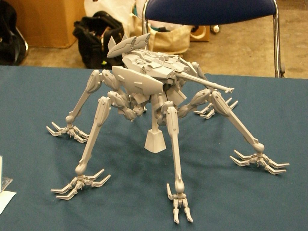
The gait guys have talked about the foot tripod for a very long time. But the truth of the matter is that it is really a HEXApod. HEXA means 6. And when the foot is properly orientated and engaged on the ground, the 5 metatarsal heads and the heel should all be engaged with the ground, truly making it an asymmetrical hexapod. In an ideal scenario, the foot would be most stable if it looked like the strange symmetrical hexapod above with the contact points equally distributed around a center point. But that is not the human foot and this version of a hexapod is far simpler and likely inferior to the foot hexapod when human locomotion is to be attempted. The human foot is engineering marvel when it works properly.  

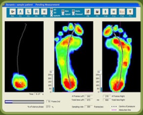
Perhaps the best example of what I mean by the foot being a HEXApod is in the pressure diagram above. In that first picture, on the right of that picture, we see multiple pressure points under the metatarsal heads of the right foot.  Minus the missing 1st metatarsal head pressure point (taken over by increased flexor hallucis longus activity represented by increased pressure at the big toe),  this pretty much confirms that the foot is not a tripod, rather a hexapod. The theory of the tripod, the 1st and 5th metatarsal heads and the heel, is only crudely accurate and honest. In this picture case, this person has increased lateral foot weight bearing (possibly why the 1st MET head pressure is absent) and possibly represented by pressure under the base of the 5 metatarsal. This is not normal for most people and if this person could get the 1st MET head down, they might even have a HEPTApod foot structure because of the 5th metatarsal base presentation (which sometimes represents peroneal muscle weakness). 

Where did we lead you astray after all these years of “tripod” talk ? We have always discussed the foot tripod. We have always discussed the imperative need to keep the limb’s plumb line forces within the area represented by the tripod.  If your forces fall more laterally within the tripod, as in this first discussed picture, one is at increased risk of inversion events and the myriad of compensations within the entire body that will occur to prevent that inversion. So again, why the tripod?  Well, it is easier to understand and it serves our clients well when it comes to finding active foot arch restoration as seen in this video of ours here.  But, the truth of the matter is that all of the metatarsal heads should be on the ground. The 2nd METatarsal is longer, the 3rd a little shorter, and the 4th and 5th even a little short than those. With the 1st MET shorter, these 5 form a kind of parabolic arc if you will. Each metatarsal head still should contact the ground and then each of those metatarsals should be further supported/anchored by their digits (toes) distally.  So the foot is actually more truly a HEXAPOD. Take the old TRIPOD theory we have always spoken about and extend a curved line beyond the forefoot bipod points (1st and 5th metatarsals) to incorporate contact points on the 2, 3 and 4th metatarsal heads. These metatarsals help to form the TRANSVERSE arch of the foot. It is this transverse arch that has given us the easily explainable foot TRIPOD because if a line is drawn between just the shorter 1st and 5th metatarsals, we do not see contact of the 2-4 metatarsal heads when we only look for pressure between these two bipod landmarks, but the obvious truth is that the 2-4 metatarsals are just longer and extend to the ground further out beyond this theoretical line drawn between the 1st and 5th MET heads.   

So, the foot is a HEXAPOD. Make no mistake about it. It is more stable than a tripod because there are more contact points inside the traditionally discussed foot tripod model. And frankly, the tripod theory is just a lie and just too fundamentally simple, unless you are an American 3 toed woodpecker.

Dr. Shawn Allen,     www.doctorallen.co

one of the gait guys

Trade Secret: Proper Calf Raise

We are selling off part of the farm here today in giving this one away.  This is an exercise we prescribe frequently.

When we rise up onto the ball of the foot, most clients and patients tend to come up and either be flush on the forefoot bipod or even a little more onto the lateral aspect of the forefoot. When asked, rarely do we hear that they have a majority of pressure over the medial half of the forefoot. This posturing tendency can lead to inversion sprains. Imagine for a minute a basketball or volleyball player, or any sport for that matter, because most involve the foot leaving the ground and returning to it.  When the foot returns to the ground, if the foot is even a slightly bit inverted (meaning they are even slightly tending towards landing on the outer half of the forefoot) an inversion sprain is at risk. This is particularly so when the lateral gastroc-soleus is weak and the peronei are weak. Forefoot valgus foot types are certainly in the risk category here and so once again we find that knowing your foot types so you can help your clients is need-to-know information.  Back to our jump and to the return to the ground from the jump, you must remember that the metatarsals are shorter and shorter as you move to the lateral foot. This means that if the load is moving laterally because of posterio-lateral compartment weakness as described above, that the sheer design of the shorter lateral metatarsals will continue to press the motion laterally. This is one of the reasons why lateral ankle strains, inversion sprains, are so frequent and repetitive (we have described the other factor in the latency of the peronei after a single inversion sprain in other blog posts here). 

So here we have our calf raise exercise. Squeezing the ball between the ankles on the up (concentric phase) and on the down phase (eccentric) with a nice isometric at the top will force the weight bearing onto the first and second metatarsals (medial forefoot) and drive the lateral compartment to press the motion medially through an isometric instead of depending so much on this compartment to protect the inversion motion through and eccentric.  We find this motor pattern terribly weak in our athletes, especially our jumping sports and certainly after inversion sprains. IF we can provide more strength to hold this medial posture during the return to the ground from a jump we can slow or delay the lateral inversion event risk.  The key to the exercise is to keep the pressure into the ball medially at all times. A wonderful additional benefit to this exercise is that the user will feel the cocontraction of the thigh adductors which further provides a medial stability effort and blends nicely with the lower abdominals.

You can see that in this case we are rehabilitating an achilles tendon repair case on the left leg.