3 things you can do NOW for patello femoral pain...


Recalcitrant PFP? In addition to your treatment regiment AND getting to THE CAUSE of the patello femoral pain (often but not always gluteus medius function), have you tried?

  • forefoot-strike running

  • increasing step rate by 10% (ie cadence)

  • "running softer"

according to this article:

"all modifications were associated with reduced patellofemoral joint force during running, compared with the participants’ normal running gait. But the modifications were also associated with immediate symptom improvement of at least one point out of 10; 62.5% of runners in the study experienced a positive symptomatic response to at least one of the gait modifications."


Easy to do, easy to implement


Esculier J-F, Bouyer LJ, Roy J-S. Immediate effects of gait retraining on symptoms and running mechanics of runners with patellofemoral pain. J Orthop Sports Phys Ther 2017;47(suppl 1):A9.


QL and Patellofemoral Pain?

photo credit: https://www.t-nation.com/training/training-disasters

photo credit: https://www.t-nation.com/training/training-disasters

"Subjects with PFP(patello femoral pain) have a higher prevalence of MTrPs (Myofascial trigger points) in bilateral GMe (gluteus medius)) and QL (quadratus lumborum) muscles. They demonstrate less hip abduction strength compared with controls, but the TPPRT (trigger point pressure release therapy, AKA ischemic compression) did not result in an increase in hip abduction strength. "

It is not surprising that when the hip is involved, the knee will be involved. As Dr. Allen often likes to say "the knee is basically in joint between 2 ball and socket joints ".

The gluteus medius and quadratus lumborum, along with the adductors are coronal plane stabilizers of the pelvis. They both have rotational components to their function as well affecting the hip directly for the former and lumbar spine for the latter. You can see our other QL articles about this here and here.

It is not much of a stretch to imagine that dysfunction of these muscles could result in trigger points and/or dysfunction of the knee (or foot for that matter ) could cause trigger points in these muscles.

Here is an article (1) examining trigger points in the gluteus medius and quadratus lumborum which, if you are familiar with Porterfield and DeRosa's work (2), are intimately linked during gait. We found it interesting that skin nick compression did not increase hip abduction strength where we find dry needling and intramuscular therapy often do.

Don't overlook these muscles and this important relationship.


  1. Roach, Sean et al.Prevalence of Myofascial Trigger Points in the Hip in Patellofemoral Pain Archives of Physical Medicine and Rehabilitation , Volume 94 , Issue 3 , 522 - 526link to free full text article: http://www.archives-pmr.org/article/S0003-9993(12)01079-9/fulltexthttp://www.archives-pmr.org/article/S0003-9993(12)01079-9/fulltext

  2. J. Porterfield, C. DeRosa (Eds.) Mechanical low back pain. 2nd ed. WB Saunders, Philadelphia; 1991


Forefoot varus and patellofemoral cartilage damage.

So you just give everyone a FOOT TRIPOD and ANKLE ROCKER exercise and think the world will all be sunshine and rainbows huh ? Beware all you movement wizards, there is far more to it !

"Knowing enough to think you're doing it right, but not enough to know you're doing it wrong." - Neil deGrasse Tyson

So your client has knee pain huh ? Look far and wide, this is a global game amigos.
"Of the 51% of limbs with forefoot varus, 91.3% had medial and 78.3% had lateral PFJ cartilage damage. . . . . this study suggest a relationship between forefoot varus and medial PFJ cartilage damage in older adults"- Lufler et al. (study link below)

*If you do not know your client has a rigid forefoot varus, and they have hip or low back pain and cannot keep their glutes activated and participating in movements, how long are you going to fail your client ? The forefoot varus may need addressed because of the excessive, abrupt degree of internal spin on the limb.

If you are truly going to treat people, people who move (yes, that means everyone !), you have to know feet and gait, BOTH. Your knowledge must go far past rudimentary knowledge of:
- high / low arch
- flat feet
- prontation and supination
- orthotics and footbeds

You will have to know your foot types, you will have to understand shoe anatomy, foot anatomy, flexible semi-flexible and rigid foot types, compensated and uncompensated foot types, and of course know how each of these responds under various loading responses. Forefoot varus will load differently in cutting sports than in sagittal locomotion such as walking and running (both of which are different even in themselves despite both being sagittal). A foot that looks like it has a flat collapsed arch has far more to it than that, and thus remedy and intervention MUST go far beyond rudimentary interventions like a "stability shoe" or orthotic. Are you practicing, coaching, training and being part of your client's solution, or are you part of the problem ? If you want to get better at this stuff, we cover it all in our several hour (very difficult for some) National Shoe Fit program (the link is on our website if you wish to become a foot/gait/shoe jediwww.thegaitguys.com). Do not be mistaken, this is far more than "shoe Fit". To know how to properly shoe fit someone, you have to know the foot types and how they compensate, load, and respond. Without this knowledge, you are just another bump in the "road of problems" without ample solutions.

- Dr. Shawn Allen, one of the gait guys

The Association of Forefoot Varus Deformity with Patellofemoral Cartilage Damage in Older Adult Cadavers. Lufler, Stefanik, Niu, Sawyer, Hoagland, Gross http://onlinelibrary.wiley.com/doi/10.1002/ar.23524/full

images courtesy of aaronswansonpt.com and studyblue.com

What are we listening to this week? The Physio edge podcast with David pope. This week they interview Kurt Lisle about anterior knee pain. Here is our synopsis:One of the things they empahasized right off the bat was that patellofemoral pain not onl…

What are we listening to this week? 

The Physio edge podcast with David pope. This week they interview Kurt Lisle about anterior knee pain. Here is our synopsis:

One of the things they empahasized right off the bat was that patellofemoral pain not only refers about the knee but also below or most importantly posterior to the knee. The fat pad had a tendency to refer more locally where is other structures can refer to other areas.

Aggravating factors for patello femoral dysfunctional pain tends to be flexion or activities involving flexion as well as compression of the knee and rest is in alleviating factor.

The fat pad pain tends to be to either side of the patellar tendon and sometimes directly under it. This can be aggravated by standing, particularly with the knee and hyperextension, which compresses the fat pad.

Patellar tendon pain tends to remain at the inferior pole of the patella on the tendon whereas patellofemoral pain has a tendency to refer more.

Physical examination pearls:

  • Patellar tendonopathy alone generally does not have effusion present where as the patellofemoral or fat pad injury may.
  • Is there pain in passive hyperextension? This generally can mean fat pad injury or potential he ligamentous injury.
  • Visually you may palpate a thickened fat pad, particularly in females.
  • Pain with passive motions generally points away from patellar tendon.
  • Dialing in as to where and when they are having their pain is an important part of the functional evaluation.

Kurt likes to do a table top examination first to ensure functional integrity of the knee before jumping right to functional tasks. His concerns are (which are valid) is the knee up to the task you’re about to ask it to do? Good advice here.
He emphasizes the need to be systematic and consistent in your examination, no matter how you examine them. Develop a routine that you follow each and every time. He recommends passively looking at the knee in extension and 90° flexion.

There is a discussion on functional movement about the hip and pelvis, knee, and foot and ankle. Emphasis is made, for example at the knee, as to “is the knee moving medially and laterally or are the femur and tibia rotating mediately or laterally” in which is precipitating the pain?

“Catching” of the patella is often due to patellofemoral pathology such as a subchondral defect, slap tear of the chondral surface, or abnormalities of the trochlea of the femur.

Advanced imaging strategies are also discussed with a brief overview of some of the things to look for.

Finally treatment strategies were discussed. It is emphasized that identifying the specific activity or change activities that’s causing any pain he’s made as well as activity modification. We were happy to hear that footwear and its role in knee as well as hepatology was discussed as well as looking at occupational contributions to the pain.

There was emphasis on exercise specificity particularly with respect to if the problem was unilateral not giving “blanket” exercises for both knees but rather concentrating on the symptomatic side.

A discussion on the use of EMG and activation patterns was also entertained with some good clinical pearls here. More marked rather than subtle changes and activation side to side seem to be more clinically significant. In other words, with respect training, can they achieve similar levels of activation on each side with a similar activity (for example isometric knee extension with the leg bent 60°).

The judicious use of tape from a functional testing standpoint was interesting. Emphasis was made that tape is not a cure and will merely a tool.

All in all and informative, concise podcast with some great clinical pearls and a nice review of the knee and patellofemoral pain.


link to PODcast: http://physioedge.com.au/pe-029-acute-knee-injuries-with-kurt-lisle/

tumblr_o39jnrMvyo1qhko2so2_1280.jpg
tumblr_o39jnrMvyo1qhko2so4_1280.jpg
tumblr_o39jnrMvyo1qhko2so5_1280.jpg
tumblr_o39jnrMvyo1qhko2so1_1280.jpg
tumblr_o39jnrMvyo1qhko2so6_1280.jpg

Medial knee pain in a skier.   Considering an orthotic?  You had better know what you are doing! 

Can you guess why this gal has pain in both knees? Especially when skinning up a hill and skiing down? 

 Take a close look at the photos above and notice the orientation of her knee with her foot. Now look at you tuberosity and drop a line straight downward.  This line should pass through or slightly lateral to the second metatarsal shaft. Can you see how it falls to the outside of this? Perhaps even between the third and fourth metatarsal?

This gal has bilateral internal tibial torsion.  When she wears a standard foot bed (creates a level surface for the right for the foot) or an orthotic without appropriate posting, it pushes her knee outside of the saggital plane. This creates abnormal patellofemoral tracking  and appears to be a major contributor to her pain. 

 You will notice that we placed a valgus post under the orthotic(  a post that is canted from lateral to medial) which pushes her knee to the midline as the first ray descends.  You can see her alignment is better with her boots on and the changes. 

 The bottom line? Know your torsions and versions.  Posting a patient like this incorrectly could result in a meniscal disaster!

Change the foot, change the knee (and vice versa). A video case of External Tibial Torsion.

Here is a perfect example of external tibial torsion. Are you treating and training people and messing with their orthotics, squat knee-foot posturing or making gait/running/jumping changes or recommendations? If you are doing all of this and you do not know about tibial torsions, then shame on you, go apologize to these people right now. You could be causing them mechanical grief. Go buy them ice cream (even if they are “paleo”), that fixes most unintentional human mistakes. 

This is a classic presentation of external tibial torsion. This is an anatomic problem, you cannot fix this intrinsically, but you can help extrinsically. You teach these people about this issue and why the foot and the knee cannot cooperate. You teach them why their feet are spun out (increased foot progression angle) while their knee tracks straight forward sagittally. You teach them why they might heel strike far laterally and why their pronation phase might be abrupt. As in this video, you teach them why they might fashionably choose to narrow the foot progression angle (foot turned in) while at the same time having to bear weight on the lateral foot (in supination to externally spin the tibia) to keep the knee tracking sagittally. You teach them why this will be impossible to do in pumps (inversion sprain ouch) and why over time this will anger many joints and tendons. You teach them that without this accommodation they will track the knee inside the sagittal plane (as seen in the video).  You teach them why they might be at greater risk of having foot prontation issue pathologies, why they might have limited internal hip rotation, why orthotics likely do not do much for them (yes, there are exceptions), why certain shoes are a challenge for them while others are magical and why over time their once beautiful arch has begun to “fall” and be less prominent as they attenuate the plantar tissues.  

As you get good with this gait and biomechanics stuff, you should readily see and understand all of the issues discussed here today in a mere flash of instant brilliance so you know what to offer your client, in understanding and remedy options. As you have seen in this video, when left to their own devices, they naturally allow the knee to find the sagittal plane in a nice forward hinge. In this posture the foot is excessively progressed outward. Again, this is because of the tibial long bone torsion. This is their anatomy, this is not functional in this case. You cannot fix this, you help them manage this, first with their awareness, then with your brilliance.  You may implement exercises and gait strategies to help them become aware of mechanical issues and how to protect the foot-ankle, the knee and the hip. You teach them why they might have a tendency towards anterior pelvis posturing or sway back type postures. You teach them why, in some cases, they choose knee hyperextension as a comfortable yet lazy stance postural habit. You teach them why some shoes are “happy” shoes for them, and why others are pure evil.

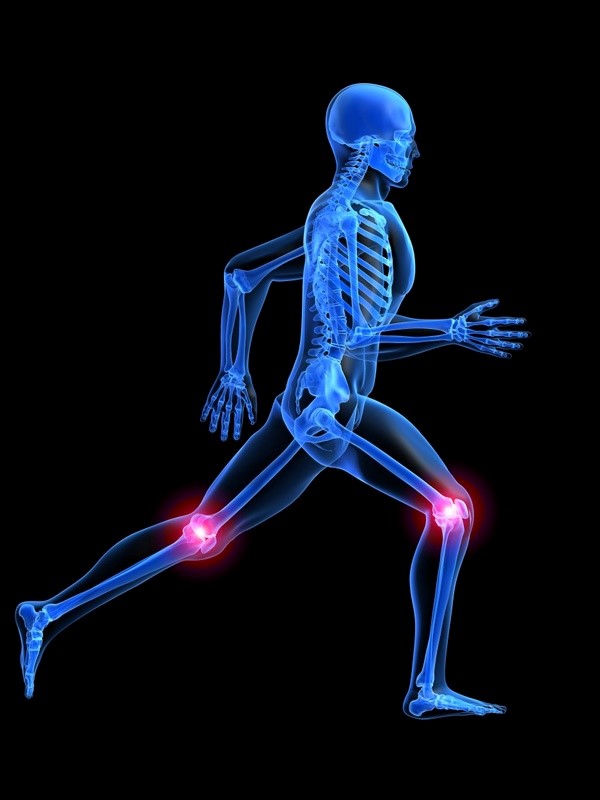
A foundational principle we teach here at The Gait Guys is that the knee is a simple hinge between two multiaxial joints on either side of the knee, the hip and the foot-ankle complex. The knee really can only flex and extend, and when the mechanics above and below are challenged the knee has little depth to its abilities to tolerate much of anything except simple sagittal hinging. You can see that the foot posturing and tibial torsion rule the roost here in this video. You should learn in time that managing this case above and below the knee is where the pot of gold is found. You will learn in time that taping the knee is often futile, yet a worthy experiment both for you and the client in the discovery process, but that a life time of taping is not logical. External tibial torsion, although affording the knee that sagittal hinge plane, can narrow its range of safe sagittal mechanics and it is up to you to  help them learn and discover that razor’s edge safely and effectively when the torsion is large.  You should also discuss with them that as they plastically tissue adapt over the years (ie. pronate more and lose more arch integrity), this razor’s edge may widen or narrow for the knee mechanics as well as the hip and foot-ankle complex.  

For your reading pleasure, a classic example of how to interrogate a safe sagittal knee progression was discussed in this blog video piece we wrote recently, linked here.

Look and you shall find, but only if you know what you are looking for.

* Please now know that you should never off the cuff tell someone to turn inwards their outwardly spun foot. But if you do, have ice cream on hand, just in case.

Need more to spin your head ? Think about whether their IT band complex is going to be functioning normally.  Oy, where is that ice cream !

Shawn Allen, one of the gait guys

Patello femoral pain? Thinking weak VMO? Think again…“Atrophy of all portions of the quadriceps muscles is present in the affected limb of people with unilateral PFP. There wasn’t any atrophy of the quadriceps in individuals with …

Patello femoral pain? Thinking weak VMO? Think again…

“Atrophy of all portions of the quadriceps muscles is present in the affected limb of people with unilateral PFP. There wasn’t any atrophy of the quadriceps in individuals with PFP compared to those without pathology. Selective atrophy of the VMO relative to the vastus lateralis wasn’t identified in persons with PFP.”

http://www.physiospot.com/research/atrophy-of-the-quadriceps-is-not-isolated-to-the-vastus-medialis-oblique-in-individuals-with-patellofemoral-pain/

Folks with patellofemoarl pain move differently. But they don’t necessarily engage their trunk differntly. We think we all knew this, but here is a study that looks at it. “Compared with the control group, the PFP group demonstrated increased ipsila…

Folks with patellofemoarl pain move differently. But they don’t necessarily engage their trunk differntly. We think we all knew this, but here is a study that looks at it. 


“Compared with the control group, the PFP group demonstrated increased ipsilateral trunk lean, hip adduction and knee abduction (p = 0.02-0.04) during single-leg squat accompanied with decreased trunk isometric strength (p = < 0.001-0.009). There was no between-group difference in trunk muscle activation. Only in the control group, ipsilateral trunk lean was significantly correlated with hip adduction (r = -0.66) and knee abduction (r = 0.49); also, the side bridge test correlated with knee abduction (r = -0.51). Differences in trunk, hip and knee biomechanics were found in people with PFP. No relationship among trunk, hip and knee biomechanics was found in the PFP group, suggesting that people with PFP show different movement patterns compared to the control group.”


Man Ther. 2015 Feb;20(1):189-93. doi: 10.1016/j.math.2014.08.013. Epub 2014 Sep 9.Trunk biomechanics and its association with hip and knee kinematics in patients with and without patellofemoral pain.Nakagawa TH1, Maciel CD2, Serrão FV3.

Foot orthoses and patellofemoral pain: frontal plane effects during running | Lower Extremity Review Magazine

We all see people with patellofemoral pain. Some of those cases may have responded to orthotic therapy. Some studies show that the effects on frontal plane kinematics are minimal (1 degree); this doesn’t mean it didn’t work, or this amount is not clinically significant. So why do they help? Perhaps it is a “timing” issue and the knee abduction moment.

“Our results are consistent with a 2003 study by Mundermann et al that compared the effects of custom orthoses (with posting, molding, or a combination of both) to flat inserts. For each orthotic condition, these authors reported a significant delay in the timing of the peak knee abduction moment. This finding may be related to the aforementioned clinical effects, as delaying the peak knee abduction moment would effectively decrease the rate of loading at the knee joint. The rate of loading has been previously implicated as a possible contributing factor in running-related overuse injuries, as runners with a history of injury have demonstrated a higher rate of loading of the vertical ground reaction force than runners with no history of running-related injury.”

This is an interesting take. If you have a few moments, give it a read:

Podcast 81: Gait, critical, pure and essential principles

This week’s show sponsors: 

www.newbalancechicago.com

www.lemsshoes.com

A. Link to our server: 

http://traffic.libsyn.com/thegaitguys/pod_81f.mp3

Direct Download: 

http://thegaitguys.libsyn.com/podcast-81-gait-critical-pure-and-essential-principles

B. iTunes link:

https://itunes.apple.com/us/podcast/the-gait-guys-podcast/id559864138

C. Gait Guys online /download store (National Shoe Fit Certification and more !) :

http://store.payloadz.com/results/results.aspx?m=80204

D. other web based Gait Guys lectures:

www.onlinece.com   type in Dr. Waerlop or Dr. Allen,  ”Biomechanics”

______________

Today’s Show notes:

Show Sponsors:
 

* Gait Guys online /download store (National Shoe Fit Certification and more !) :

http://store.payloadz.com/results/results.aspx?m=80204

* Other web based Gait Guys lectures:

www.onlinece.com   type in Dr. Waerlop or Dr. Allen,  ”Biomechanics”

 
Show Notes and links:
 
Forget Cheetah Blades. This Prosthetic Socket Is a Real Breakthrough
http://www.wired.com/2014/10/forget-cheetah-blades-prosthetic-socket-real-breakthrough
 
Rebuilding and Regenerating Damaged Knees: The Future Has Arrived!
http://www.huffingtonpost.com/nicholas-dinubile-md/rebuilding-and-regenerati_b_6043374.html
 
the foot gym:
 
From a reader:
Thanks for sharing all the great information over the years. I would like to pose to you some simple questions. How do you decide what area/s are relevant to the issue a patient presents? How do you decide what is “normal” given anatomical variations, history of injuries, torsion’s, etc., and if pain is present, why would you address biomechanics, since pain is a neurological phenomenon not a biomechanical phenomenon?
This may not be that simple but would like to hear what you have to say on these topics.
Thank you,
Joe 
 
COMPARISON OF ISOMETRIC ANKLE STRENGTH BETWEEN FEMALES WITH AND WITHOUT PATELLOFEMORAL PAIN SYNDROME
http://www.ncbi.nlm.nih.gov/pmc/articles/PMC4196327/
 
the drawbacks of technology

Video case: The King’s Preference: Short and Sweet. A quick and easy case demonstrating the patellar tracking struggles with external tibial torsion.

Our favorite functional evaluation piece of equipment as well as our favorite piece of therapy equipment is the Total Gym.  Here we clearly demonstrate, to us and the client, in partial weight bearing load, the effects of external tibial torsion.  

Remember, the knee is sort of the King of all joints when it comes to the lower extremity.  The knee is a sagittal plane hinge, and so all it wants to do is hinge forward, freely without binding from deficits at the hip or knee. But we cannot ignore the simple fact that pre-pubescent kids the long bone derotation process is still undergoing, and in adults the process may have been corrupted or insufficient.  

In this case it should be obvious that the knee is sagittal and free to hinge when the foot is at a large foot progression angle.  This allows the knee to hinge cleanly. But when the foot is corrected to the sagittal plane, as you see in the second half of the video, the knee tracks inward and this can cause patellofemoral pain syndromes, swelling, challenges to the menisci (and possible eventual tears) and challenges to the ACL and other accessory restraints.  Additionally, this medial drift is a longer and more difficult challenge to the eccentric phase external rotators such as the gluteus maximius not to mention many of the other muscles and their optimal function.  

So, the next time you see a large foot progression angle in a client or in their walk (duck footed if you will) try to resist the natural urge to tell them to corrrect the foot angle. They are likely doing it to keep the King happy.  And furthermore, be careful on your coaching recommendations during squats, olympic lifts, lunges and running.  Just because you do not like the way the foot looks doesn’t mean you should antagonize the King of joints.  

External tibial torsion, its not something you want to see, but when you do see it, you have to know its degree, its effects at the knee, hip and foot as well as how it might impact hip extension, pelvic neutrality, foot strike, foot type, toe off and so many other aspects.

Whoever said gait analysis was easy was a liar. And if all they use is a video camera and fancy analysis software they have show up with only part of the team. And if they said they were an expert  in gait only a few years into practice, you had better also look for a jester’s hat somewhere hiding in the corner. After all, the King would want to know !

Shawn and Ivo, your court jesters for the last 3+ years.  Maybe we will get a promotion from the King someday soon !

More Proof that the Cross Over Gait has Pathologic Issues for Runners / Athletes.

We have referenced below yet another article in our 2 year long soap box rant that the cross over gait has many negatives to it.  Two weeks ago we discussed the issues in greater depth in podcast # 23 (link: http://thegaitguys.tumblr.com/post/43424418001/podcast-23-neurology-of-walking-babies-dialogues-on) and further in a most recent blog post here (link: http://thegaitguys.tumblr.com/post/44060333371/step-width-alters-iliotibial-band-strain-during).

By this point pretty much everyone should be aware that pelvis width and femoral shaft angle orientation (Q-angle) parlays consistently into knee posturing and thus patellar tracking.  Loosely it goes to say a wider pelvis often makes for a knee tracking challenged environment.  But today’s reference article takes this a little deeper.

Running mechanics always have to be approached from above the knee and below. If the foot collapses too far inwards the internal spin put on the tibia will drag the knee inwards and generate a mal-tracking environment.  And from above, if the gluteal muscles are underperforming they cannot assist in holding the femur in sufficient abduction and external rotation to prevent excessive internal spin from above, thus also enabling a mal-tracking environment from developing.  These are well established theories with plenty of research and years to back them up.  The verbiage “proximal control for distal control” holds. Or, “proximal stability for (proper) distal mobility” also holds true but one needs to never forget about the critical importance of the far distal (foot/ankle) foundational support. 

In today’s study from 2012 there is really nothing earth shatterning to most of our readers but we wanted to again bring these thoughts are results to you and keep the cross over gait in your ever-present mind.  The conclusions of this Harvard study were predictable, that being:

“the finding of greater hip adduction in female runners who develop PFP is in agreement with previous cross sectional studies. These results suggest that runners who develop PFP utilize a different proximal neuromuscular control strategy than those who remain healthy. Injury prevention and treatment strategies should consider addressing these altered hip mechanics.”

So the study eludes to the fact that not only is it about the anatomy of the parts but also about the functional control of the parts. Without adequate control from above and support from below the knee, it will be difficult to control a largely uni-planar joint (the sagittal flexion/extension of the knee hinge) when the support of a multi-planar joint complex (foot/ankle) from below is insufficient and the control of a multi-planar joint complex above the knee (hip/pelvis) is insufficient.  When one or especially both are compromised the knee will be compromised. It may take weeks or months or even longer for the process to render joint change or pain but without sufficient biomechanics the system is likely to fail. And further more, one needs to realize that shoes and orthotics often are an incomplete (and very often an insufficient and inadequate) remedy.  One must “earn it to own it”. 

If you find you are new to our work and want to catch up on the Cross Over gait topics we have covered previously, try starting here (link: http://thegaitguys.tumblr.com/search/cross+over) and here (link: http://thegaitguys.tumblr.com/search/cross+over+gait).  We are likely to continue to build on this disfunctional paradigm.

Shawn and Ivo
The Gait Guys

Reference:
Med Sci Sports Exerc. 2012 Dec 27. [Epub ahead of print] Prospective Evidence for a Hip Etiology in Patellofemoral Pain. Noehren B, Hamill J, Davis I. Source

1Division of Physical Therapy, University of Kentucky, Lexington, KY 2Department of Exercise Science, University of Massachusetts, Amherst, MA 3Spaulding National Running Center, Harvard University, Cambridge, MA.

Abstract PURPOSE:

Patellofemoral pain (PFP) is the leading cause of knee pain in runners. Proximal and distal running mechanics have been linked to the development of PFP. However, the lack of prospective studies limits establishing a causal relationship of these mechanics to PFP. The purpose of this study was to prospectively compare running mechanics in a group of female runners who went on to develop PFP compared to healthy controls. It was hypothesized that runners who go on to develop PFP would exhibit greater hip adduction, hip internal rotation, and greater rear foot eversion.

CONCLUSIONS:

The finding of greater hip adduction in female runners who develop PFP is in agreement with previous cross sectional studies. These results suggest that runners who develop PFP utilize a different proximal neuromuscular control strategy than those who remain healthy. Injury prevention and treatment strategies should consider addressing these altered hip mechanics.

What have we here?

Take a look at the tibial tuberosity and then where you think the 2nd metatarsal head would be. What do you see? The 2nd metatarsal is lateral to the tibial tuberosity. You are looking at external tibial torsion.

Lets see how this external tibail torsion behaves during a knee bend on a total gym. Observe the medial drift of the knee during weight bearing knee flexion. 

In external tibial torsion there is an external torsion or a “twist” along the length of the tibia (diaphysis or long section) (need a review? click here). This occurs in this example to the degree that the ankle joint (mortise joint) can no longer cooperate with sagittal knee joint.  When taking a client with external tibial torsion and pre-postioning their foot in a relatively acceptable/normal foot progression angle as seen here, there is a conflict at the knee, meaning that the knee cannot hinge forward in its usual sagittal plane. In this case with the foot progression angle smaller than what this client would posture the foot, you an see that as they bend the knee the knee is forced to drift medially and as soon as the heel is unloaded a pure “adductory twist” is noted (you can see the heel jump medially in an attempt to find a more tolerable sagittal knee bend).

Are you looking for torsions of the lower limb in your clients ?

Are you forcing them into foot postures that look better to  you but that which are conflicting to your clients given body mechanics ?  Remember, telling someone to turn their foot in or out because it doesn’t appear correct to your eyes can significantly impair either local or global joints , and often both. Torsions can occur in the talus, the tibia and the femur.

Furthermore, torsions can have an impact on foot posturing at foot strike and affect the limbs loading response, from foot to core and even arm swing can be altered.  Letting your foot fall naturally beneath your body does not mean that you have the clean anatomy to do so without a short term or long term cost. 

Want more on torsion and versions ?  Type the words into the search box on our blog. We have plenty of good info for you.

Shawn and Ivo, The Gait Guys

Hip rotation and knee pain. What we have been saying.

We have been pounding the floor on this topic on and off for the last 6 months it seems.  Lack of internal hip rotation or too much internal hip rotation ……. both abnormal hip kinematics, is a result of reduced hip-muscle performance as opposed to structural issues of the hip (anteversion, retroversion etc).

If you are not assessing for impaired hip muscle function in your knee patients, you could be missing the boat …….. and it is a big boat…… Titanic in size.

Don’t be like so many others and be tunnel visioned when you have a knee patient, expand your vision, at least to the hip and foot. 

We are……. The Gait Guys……. Shawn and Ivo

________________________________________________________________________________

Am J Sports Med. 2009 Mar;37(3):579-87. Epub 2008 Dec 19.

Predictors of hip internal rotation during running: an evaluation of hip strength and femoral structure in women with and without patellofemoral pain.

Souza RB, Powers CM.

Musculoskeletal and Quantitative Imaging Research Laboratory, Department of Radiology and Biomedical Engineering, University of California, San Francisco, California, USA.