A bit about the QL...


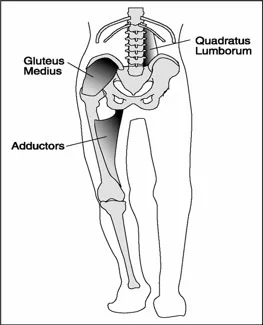
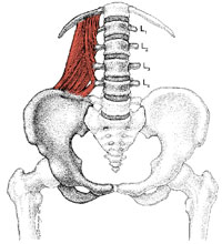
As we have said in previous posts,  though they can’t act independently we like to think to think of the QL as having two divisions. The lower division arises from the medial portion of the iliac crest and adjacent iliolumbar ligament and inserts onto the transverse processes of the lumbar vertebrae, in the coronal plane from lateral to medial and in the saggital plane from posterior to anterior. The upper division arises from the lumbar transverse processes of the upper 4 lumbar vertebrae and insert into the inferior border of the 12th rib, running in the coronal plane from medial to lateral and in the saggital plane from anterior to posterior; about half of the fascicles of this second division act on the twelfth rib and the rest act on the lumbar spine.

The QL is primarily a coronal plane stabilizer causing lateral bending to the ipsilateral side when the foot is planted as well as posterior rotation of the lumbar spine on the weight bearing side.   When acting unilaterally without the ipsilateral foot fixed on the ground, it can raise the ilia on the side of contraction. It is active during single limb support during stance phase of gait on the contralateral side (along with the external oblique) to elevate the ilium. This is coupled with the ipsilateral anterior fibers of the gluteus medius and minimus pulling the iliac crest toward the stable femur. Sahrmann states “the QL is optimally situated to provide control of lateral flexion to the opposite side via its eccentric contraction to provide control of the return from lateral flexion via its concentric contraction. The muscle is also positioned to play a role in the rotation that occurs between the pelvis and spine during walking”.

Acting bilaterally, it extends the lumbar spine, deepening the lordosis and acting to limit anterior shear of the vertebral bodies.

It is also able to stabilize the 12th rib during forced expiration, thus acting as an accessory muscle of respiration. This fixation is important when we need to superimpose pelvic movements upon it. Furthermore, it increased activation in response to increasing compression in static upright standing postures.

Here is a video of a low back screen we often use

QL and Patellofemoral Pain?

photo credit: https://www.t-nation.com/training/training-disasters

photo credit: https://www.t-nation.com/training/training-disasters

"Subjects with PFP(patello femoral pain) have a higher prevalence of MTrPs (Myofascial trigger points) in bilateral GMe (gluteus medius)) and QL (quadratus lumborum) muscles. They demonstrate less hip abduction strength compared with controls, but the TPPRT (trigger point pressure release therapy, AKA ischemic compression) did not result in an increase in hip abduction strength. "

It is not surprising that when the hip is involved, the knee will be involved. As Dr. Allen often likes to say "the knee is basically in joint between 2 ball and socket joints ".

The gluteus medius and quadratus lumborum, along with the adductors are coronal plane stabilizers of the pelvis. They both have rotational components to their function as well affecting the hip directly for the former and lumbar spine for the latter. You can see our other QL articles about this here and here.

It is not much of a stretch to imagine that dysfunction of these muscles could result in trigger points and/or dysfunction of the knee (or foot for that matter ) could cause trigger points in these muscles.

Here is an article (1) examining trigger points in the gluteus medius and quadratus lumborum which, if you are familiar with Porterfield and DeRosa's work (2), are intimately linked during gait. We found it interesting that skin nick compression did not increase hip abduction strength where we find dry needling and intramuscular therapy often do.

Don't overlook these muscles and this important relationship.


  1. Roach, Sean et al.Prevalence of Myofascial Trigger Points in the Hip in Patellofemoral Pain Archives of Physical Medicine and Rehabilitation , Volume 94 , Issue 3 , 522 - 526link to free full text article: http://www.archives-pmr.org/article/S0003-9993(12)01079-9/fulltexthttp://www.archives-pmr.org/article/S0003-9993(12)01079-9/fulltext

  2. J. Porterfield, C. DeRosa (Eds.) Mechanical low back pain. 2nd ed. WB Saunders, Philadelphia; 1991


Not quite the QL, but close....

Screen Shot 2017-08-28 at 10.13.40 AM.png

We all see folks with low back pain and gait abnormalities. It is active during single limb support during stance phase of gait on the contralateral side (along with the external oblique) to elevate the ilium. This is coupled with the ipsilateral anterior fibers of the gluteus medius and minimus pulling the iliac crest toward the stable femur.

We found this FREE FULL TEXT while doing some quadratus lumborum research. It reminds us about things like scleratogenous pain (pain arising from tissues of like embryological origin with a common nerve innervation, like tendon, bone, muscle, etc) and other triggers for low back pain. We have needled this ligament with good result. Remember that this is an individual ligament making up a portion of the middle layer of the thoracolumbar fascia, and is not an aponeurosis of the lumbocostal fibers of the quadratus lumborum.


https://www.ncbi.nlm.nih.gov/pmc/articles/PMC5226660/

photo from: https://musculoskeletalkey.com/treatment-of-the-patient-with-chronic-pain/

Go ahead and try this at home.remember last mondays post? (if not, click here). Here is one way of telling whether your (or someone else’s) vestibular system is working. It will also give you an idea of how some people compensate. Ready?
Stand up (b…

Go ahead and try this at home.

remember last mondays post? (if not, click here). Here is one way of telling whether your (or someone else’s) vestibular system is working. It will also give you an idea of how some people compensate.

Ready?

  • Stand up (barefoot or shoes does not matter).
  • place your hands resting on the top of your hips with your thumbs to the back (like your Mom used to, when you were in trouble). Your thumbs should be resting on your quadratus lumborum (QL) muscle.
  • tilt your HEAD to the LEFT
  • you should feel the muscle (ie the QL) under your RIGHT thumb contract
  • come back upright


repeat, but this time lean your BODY to the LEFT

  • same thing right? Now check the other side.


Everything OK? Everything fire as it should?

Now lets add another dimension.

  • slide your fingers down so they are just below the crest of the hip, resting above the greater trochanter (the bump on the side of your upper thigh). This should place your fingers on the middle fibers of the gluteus medius.
  • tilt your head (or body ) to the LEFT.
  • You should feel the LEFT gluteus medius and the RIGHT QL contract. These muscles should be paired neurologically. When walking, during stance phase on the LEFT: the LEFT gluteus medius helps to maintain the pelvis level, while the RIGHT QL, assists in hiking the RIGHT side.


If everything works OK, then your vestibulospinal spinal system is intact and your QL and gluteus medius seem to be firing and appropriately paired. If not? That is the subject for another post.

The Gait Guys. Helping you to understand the concepts of WHY compensations occur.

tumblr_lohzpbv2Zj1qhko2so1_500.png
tumblr_lohzpbv2Zj1qhko2so2_250.jpg

The Mighty Quadratus: Part 2  The Quadratus and Gait

Acting unilaterally without the ipsilateral foot fixed on the ground, it can raise the ilia on the side of contraction (as in a pull up or side bend on a Roman chair). The quadratus lumborum was more active than other muscles during isometric side support postures where the body is held horizontally almost parallel to the floor as the subjects supported themselves on one elbow on the floor together with both feet. 

It is active during single limb support during stance phase of gait on the contralateral side (along with the external oblique) to elevate the ilium. This is coupled with the ipsilateral anterior fibers of the gluteus medius and minimus pulling the iliac crest toward the stable femur.

Sahrmann states “the QL is optimally situated to provide control of lateral flexion to the opposite side via its eccentric contraction to provide control of the return from lateral flexion via its concentric contraction. The muscle is also positioned to play a role in the rotation that occurs between the pelvis and spine during walking”. This makes you really think about the interplay of this muscle, and another stance phase stabilizer, the psoas major, which attaches opposite the QL on the anterior aspect of the vertebral body, IN FRONT of the transverse process.

Acting bilaterally, it extends the lumbar spine, deepening the lordosis and acting to limit anterior shear of the vertebral bodies.

It is able to stabilize the 12th rib during forced expiration, thus acting as an accessory muscle of respiration. This fixation is important when we need to superimpose pelvic movements upon it. Furthermore, it increased activation in response to increasing compression in static upright standing postures.

Bottom Line?:

Think of the QL, especially during gait abnormalities or recalcitrant low back pain. The more it is stressed, the more it is activated. If someone had mild weakness of the stance leg gluteus medius, it may be called into play to pick up some of the slack. Expect to see increased activity paraspinally, with particular attention paid to the 12 rib attachments.

In our flexor dominant society, the QL may play a role in generating unilateral shear forces on the lumbar spine (along with the ipsilateral psoas), especially in individuals with poor ankle rocker or decreased hip extension.

The QL: it’s not only for breakfast anymore…..

We still are…The Gait guys