Sometimes you may need to put the cart before the horse...The knees, the glutes and reverse engineering ?

Footnotes 7 - Black and Red.jpg

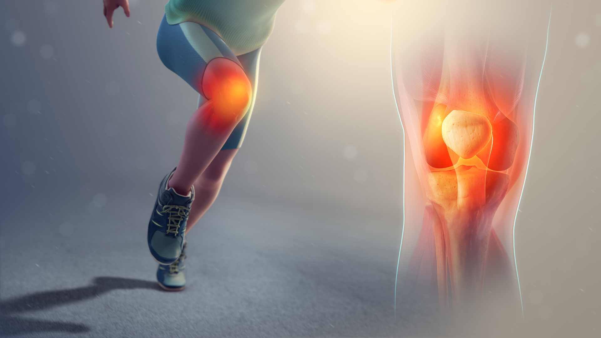
We have talked about looking at things “from the bottom” up in the past, so we can understand things like why the vastus medialis is an external rotator in closed chain as are the semi membranosis and tendinosis. Perhaps we need to think more about this traveling proximally, where the knee effects the glutes. We found this paper looking at women with patello femoral problems and gluteal inhibition. Prospective studies have not found gluteal weakness to be a risk factor for patello femoral problems, but perhaps it is the other way around and patello femoral problems are a risk factor for gluteal weakness? It makes sense, especially if you consider the vastus lateralis like we talk about here and here.

“We hypothesize that muscle inhibition is present in the gluteal muscles of females with PFP compared to healthy controls and it is associated with both decreased subjective function and longer duration of symptoms.”

Dr Ivo Waerlop, one of The Gait Guys

Glaviano NRBazett-Jones DMNorte G. Gluteal muscle inhibition: Consequences of patellofemoral pain? Med Hypotheses. 2019 May;126:9-14. doi: 10.1016/j.mehy.2019.02.046. Epub 2019 Feb 27.

#gait #foot #patellofemoralpain #PFP #quadriceps #thegaitguys #glutes #gluteal muscles

All that creaks may not be pathological...

image source: https://commons.wikimedia.org/wiki/File:Runners-knee_SAG.jpg

image source: https://commons.wikimedia.org/wiki/File:Runners-knee_SAG.jpg

Gal with creaky knees? Patellar crepitus? Does all that noise mean something?

Well, it means that knee function is suboptimal and more than likely, there is abnormal patellar tracking. But is that clinically significant? The answer is ....maybe.

This study (1) looked at over 300 women, about 1/2 with patellofemoral pain and half without looking at the following outcomes: 

  • the knee crepitis test
  • anterior knee pain scale
  • self reported knee pain in the last month
  • knee pain after 10 squats 
  • knee pain after climbing 10 stairs

They found that if you had patello femoral pain, you were 4 times more likely to have crepitus than not, but there was no correlation of crepitus with  Knee crepitus had no relationship with function, physical activity level , worst pain, pain climbing stairs or pain squatting. 

We would have loved to have seen any correlation in this group with knee valgus angles (i.e. "Q" angles 2 ) and how much tibial or femoral torsion was present (as these things change pressure and contact area 3), but that will hopefully be found in the literature elsewhere. 


1. , Pazzinatto MFPriore LBDFerreira ASBriani RVFerrari DBazett-Jones DAzevedo FM. Knee crepitus is prevalent in women with patellofemoral pain, but is not related with function, physical activity and pain. Phys Ther Sport. 2018 Sep;33:7-11. doi: 10.1016/j.ptsp.2018.06.002. Epub 2018 Jun 6.

2. Emami MJ1, Ghahramani MHAbdinejad FNamazi H. Q-angle: an invaluable parameter for evaluation of anterior knee pain. Arch Iran Med. 2007 Jan;10(1):24-6.

3. Thay Q. Lee, PhD, Garrett Morris, BS, Rick P. Csintalan, MDThe Influence of Tibial and Femoral Rotation on Patellofemoral Contact Area and Pressure Orthop Sports Phys Ther 2003;33:686-693.

wider, flatter, less mobile feet

Screen Shot 2018-05-03 at 6.59.44 AM.png

If you have patella femoral pain, the older you get, the wider (probably for increased proprioception), flatter (possibly due to loss of intrinsic strength and extensor tone) and less mobile (for stability) you feet become. 


"This study observed that in individuals with PFP, those aged 40–50 years had less foot mobility than younger adults aged 18–29 years, as evidenced by measures of midfoot height mobility and foot mobility magnitude. These differences represented a moderate effect size, and exceed the intra-rater minimal detectable change (MDC 95%) associated with these measures (midfoot height mobility 2 mm; foot mobility magnitude 3.1 mm). The differences between age groups were specific to both midfoot height mobility and foot mobility magnitude; however, there were no differences in midfoot width mobility."


Tan JM, Crossley KM, Vicenzino B, Menz HB, Munteanu SE, Collins NJ. Age-related differences in foot mobility in individuals with patellofemoral pain. Journal of Foot and Ankle Research. 2018;11:5. doi:10.1186/s13047-018-0249-2.

free full text: https://www.ncbi.nlm.nih.gov/pmc/articles/PMC5815185/


Unless you have ownership....

Compliance is often the issue ...especially in younger folks

Just say no to the exercise video. You need:

  • understanding on the patients part of the pathology and the importance of the rehab
  • buy in on the patients part
  • a way to monitor progress with objective outcomes

a nice review article in LER, full text here

additionally, this was covered in a great PODcast by David Pope here: http://physioedge.com.au/physio-edge-039-patellofemoral-pain-adolescents-dr-michael-rathleff/

What are we listening to this week? The Physio edge podcast with David pope. This week they interview Kurt Lisle about anterior knee pain. Here is our synopsis:One of the things they empahasized right off the bat was that patellofemoral pain not onl…

What are we listening to this week? 

The Physio edge podcast with David pope. This week they interview Kurt Lisle about anterior knee pain. Here is our synopsis:

One of the things they empahasized right off the bat was that patellofemoral pain not only refers about the knee but also below or most importantly posterior to the knee. The fat pad had a tendency to refer more locally where is other structures can refer to other areas.

Aggravating factors for patello femoral dysfunctional pain tends to be flexion or activities involving flexion as well as compression of the knee and rest is in alleviating factor.

The fat pad pain tends to be to either side of the patellar tendon and sometimes directly under it. This can be aggravated by standing, particularly with the knee and hyperextension, which compresses the fat pad.

Patellar tendon pain tends to remain at the inferior pole of the patella on the tendon whereas patellofemoral pain has a tendency to refer more.

Physical examination pearls:

  • Patellar tendonopathy alone generally does not have effusion present where as the patellofemoral or fat pad injury may.
  • Is there pain in passive hyperextension? This generally can mean fat pad injury or potential he ligamentous injury.
  • Visually you may palpate a thickened fat pad, particularly in females.
  • Pain with passive motions generally points away from patellar tendon.
  • Dialing in as to where and when they are having their pain is an important part of the functional evaluation.

Kurt likes to do a table top examination first to ensure functional integrity of the knee before jumping right to functional tasks. His concerns are (which are valid) is the knee up to the task you’re about to ask it to do? Good advice here.
He emphasizes the need to be systematic and consistent in your examination, no matter how you examine them. Develop a routine that you follow each and every time. He recommends passively looking at the knee in extension and 90° flexion.

There is a discussion on functional movement about the hip and pelvis, knee, and foot and ankle. Emphasis is made, for example at the knee, as to “is the knee moving medially and laterally or are the femur and tibia rotating mediately or laterally” in which is precipitating the pain?

“Catching” of the patella is often due to patellofemoral pathology such as a subchondral defect, slap tear of the chondral surface, or abnormalities of the trochlea of the femur.

Advanced imaging strategies are also discussed with a brief overview of some of the things to look for.

Finally treatment strategies were discussed. It is emphasized that identifying the specific activity or change activities that’s causing any pain he’s made as well as activity modification. We were happy to hear that footwear and its role in knee as well as hepatology was discussed as well as looking at occupational contributions to the pain.

There was emphasis on exercise specificity particularly with respect to if the problem was unilateral not giving “blanket” exercises for both knees but rather concentrating on the symptomatic side.

A discussion on the use of EMG and activation patterns was also entertained with some good clinical pearls here. More marked rather than subtle changes and activation side to side seem to be more clinically significant. In other words, with respect training, can they achieve similar levels of activation on each side with a similar activity (for example isometric knee extension with the leg bent 60°).

The judicious use of tape from a functional testing standpoint was interesting. Emphasis was made that tape is not a cure and will merely a tool.

All in all and informative, concise podcast with some great clinical pearls and a nice review of the knee and patellofemoral pain.


link to PODcast: http://physioedge.com.au/pe-029-acute-knee-injuries-with-kurt-lisle/

tumblr_o39jnrMvyo1qhko2so2_1280.jpg
tumblr_o39jnrMvyo1qhko2so4_1280.jpg
tumblr_o39jnrMvyo1qhko2so5_1280.jpg
tumblr_o39jnrMvyo1qhko2so1_1280.jpg
tumblr_o39jnrMvyo1qhko2so6_1280.jpg

Medial knee pain in a skier.   Considering an orthotic?  You had better know what you are doing! 

Can you guess why this gal has pain in both knees? Especially when skinning up a hill and skiing down? 

 Take a close look at the photos above and notice the orientation of her knee with her foot. Now look at you tuberosity and drop a line straight downward.  This line should pass through or slightly lateral to the second metatarsal shaft. Can you see how it falls to the outside of this? Perhaps even between the third and fourth metatarsal?

This gal has bilateral internal tibial torsion.  When she wears a standard foot bed (creates a level surface for the right for the foot) or an orthotic without appropriate posting, it pushes her knee outside of the saggital plane. This creates abnormal patellofemoral tracking  and appears to be a major contributor to her pain. 

 You will notice that we placed a valgus post under the orthotic(  a post that is canted from lateral to medial) which pushes her knee to the midline as the first ray descends.  You can see her alignment is better with her boots on and the changes. 

 The bottom line? Know your torsions and versions.  Posting a patient like this incorrectly could result in a meniscal disaster!