Why alignment of the big toe is so critical to gait, posture, stabilization motor patterns and running.
There are two ways of thinking about the arch of the foot when it comes to competent height. One perspective is to passively jack up the arch with a device such as an orthotic, a choice that we propose should always be your last option, or better yet to access the extrinsic and intrinsic muscles of the foot (as shown in this video) to compress the legs of the foot tripod and lift the arch dynamically. Here today we DO NOT discuss the absolute critical second strategy of lifting the arch via the extensors as you have seen in our “tripod exercise video” (link here) but we assure you that regaining extensor skill is an absolute critical skill for normal arch integrity and function. We like to say that there are two scenarios going on to regain a normal competent arch (and that does not necessarily mean a high arch, a low arch can also be competent….. it is about function and less about form): one scenario is to hydraulically lift the arch from below and the other scenario is to utilize a crane-like effect to lift the arch from above. When you combine the two, you restore the arch function. In those with a flat flexible incompetent foot you can often regain normal alignment and function. But remember, you have to get to the client before the deforming forces are significant enough and have been present long enough that the normal anatomical alignments are no longer possible. For example, a hallux valgus with a large bunion (this person will never get to the abductor hallucis sufficiently) or a progressively collapsed arch that is progressively becoming rigid or semi-rigid.
Think about these concepts today as you watch your clients walk, run or exercise. And then consider this study below on the critical importance of the abductor hallucis muscle after watching our old video of Dr. Allen’s competent foot.
CONCLUSIONS AND CLINICAL RELEVANCE:
The abductor hallucis muscle acts as a dynamic elevator of the arch. This muscle is often overlooked, poorly understood, and most certainly rarely addressed. Understanding this muscle and its mechanics may change the way we understand and treat pes planus, posterior tibial tendon dysfunction, hallux valgus, and many other issues that lead to a challenge of the arch, effective and efficient gait. Furthermore, its dysfunction and lead to many aberrant movement and stabilization strategies more proximally into the kinetic chains.
*From the article referenced below, “Most studies of degenerative flatfoot have focused on the posterior tibial muscle, an extrinsic muscle of the foot. However, there is evidence that the intrinsic muscles, in particular the abductor hallucis (ABH), are active during late stance and toe-off phases of gait.“
We hope that this article, and the video above, will bring your focus back to the foot and to gait for when the foot and gait are aberrant most proximal dynamic stabilization patterns of the body are merely strategic compensations.
Study RESULTS:
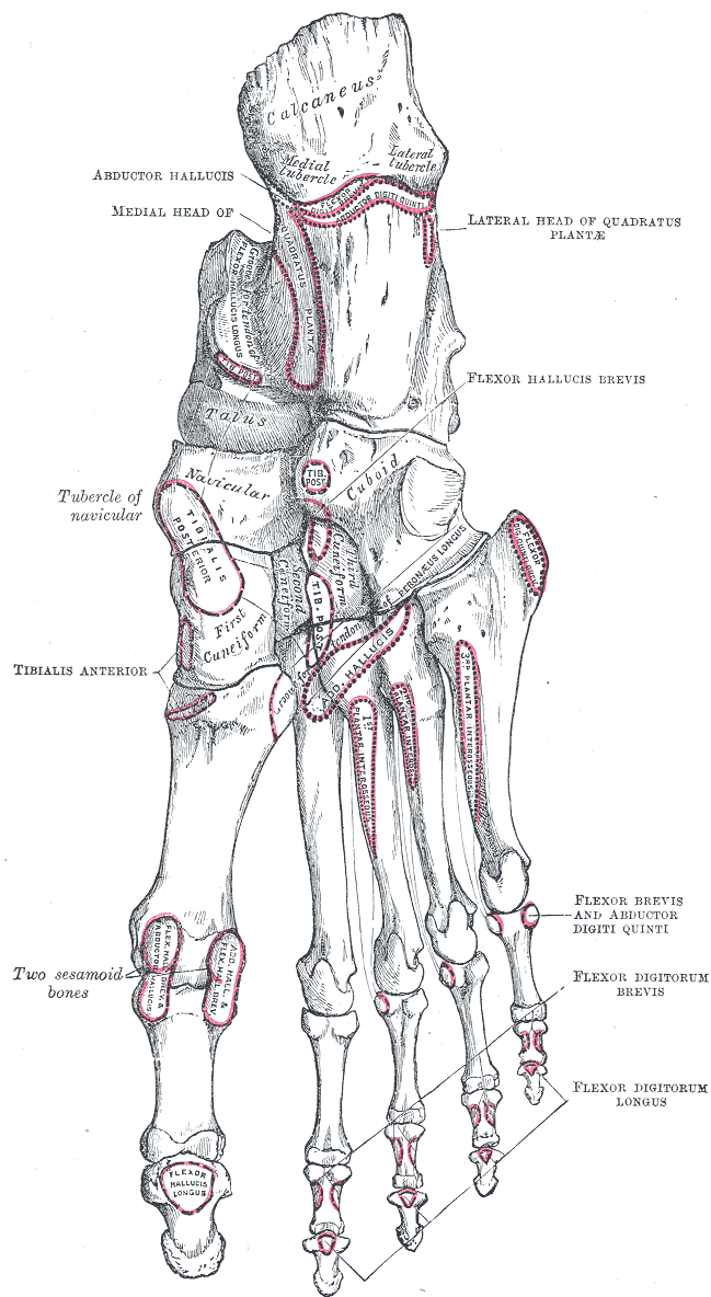
All eight specimens showed an origin from the posteromedial calcaneus and an insertion at the tibial sesamoid. All specimens also demonstrated a fascial sling in the hindfoot, lifting the abductor hallucis muscle to give it an inverted ‘V’ shaped configuration. Simulated contraction of the abductor hallucis muscle caused flexion and supination of the first metatarsal, inversion of the calcaneus, and external rotation of the tibia, consistent with elevation of the arch.
http://www.ncbi.nlm.nih.gov/pubmed/17559771
Foot Ankle Int. 2007 May;28(5):617-20.
Influence of the abductor hallucis muscle on the medial arch of the foot: a kinematic and anatomical cadaver study.
Wong YS. Island Sports Medicine & Surgery, Island Orthopaedic Group, #02-16 Gleneagles Medical Centre, 6 Napier Road, Singapore, 258499, Singapore.


