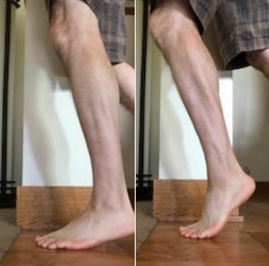
What does progressive weakness of the posterior compartment look like?

Look at this video carefully and what do you notice? Can you see the progressive dip in the left heel as time goes on while toe walking? This is a cardinal sign of lack of endurance in the posterior compartment, in this patient’s case tibialis posterior. Your differential, in addition to lack of type one muscle fibers, insufficient vascularity or mitochondria for whatever reason would be circulatory problems as well as conditions causing progressive motor weakness like myasthenia gravis.

Fatigue testing is very important because a lot of times the problem doesn’t come out till the person reaches say a half an hour, an hour or sometimes even many miles into the run or ride. Our job as clinicians is to try to diagnose the problem to the best of our abilities. Our job also is to “exploit their weaknesses” rather than “extol their virtues”. 

If you’re getting somebody with posterior calf pain or a foot drop, or maybe somebody who gets worse over time, consider fatigue testing.

Top end heel raises. The top end might matter.

Screen Shot 2019-04-19 at 6.34.09 PM.png

Thought experiment . . .
If top end posterior compartment (loosely, the calf complex) strength is lacking, then heel rise may not be optimized to transfer body mass forward sufficiently and effectively.
This lack of forward progression, fails to move the body mass sufficiently forward enough to reduce the external moment arms and optimize the internal moment arms to take maximal advantage of the calf complex (I am talking about moment arms between the grounded 1st MTP joint and ankle mortise & ankle mortise and achilles tendon).
These are rough thoughts today gang, letting you inside our heads and how we juggle multiple parameters when we are struggling to solve a client's problems.

In the lower heel rise photo, The body mass does not progress forward enough over the grounded first MTP joint at the big toe (during gait, the heel doesn’t just rise up, the axis of the ankle joint moves both up and forward).
In this case, the foot may not be fully rigid in a supinated position to benefit from joint closed-packed positions. Thus, the foot may be more pliable and one might suppose that if not adequately supinated, they are inadequately still too much relatively pronated. This might put more load into the tibialis posterior and other soft tissue mechanical loading scenarios that are less optimally suited to do this job. Over time, might this lead to pathology? Likely.
Thus, when running on a weaker posterior mechanism (often found unilaterally) the higher up posterior chains might be overburdened, the tendon loads and loading response of the achilles, tibialis posterior, and long flexors will be most likely altered, likely negatively, the naturally occurring foot locking mechanisms might be less optimal than desired, subtalar and forefoot loading might be premature (ie. sesamoid malpositioning for one, as a simple example), etc etc. Loading a foot(the mid and forefoot) into heel rise that is still somewhat pronated creates a different moment arm around the subtalar joint axis (that moves through the 1st metatarsal), than a foot that is more supinated.

Now, put these ideas into the 2 photos from yesterday where one might be loading the forefoot laterally or more medially, and now make the top end strength more in one of those scenarios. Is it any wonder why so many struggle with posteiror mechanism tendonopathies ? There are so many parameters to consider and examine. And, if not examined in great detail, the key lacking parameter can be missed.
Hence, just forcing calf strength loading is too simple a solution, there is a needle in that haystack that upset the client's apple cart, it is the job of the clinician to find it and remedy it.

Today, looking into the research and finding some interesting things that are spurring some thoughts.

Shawn Allen, one of the gait guys

What do you know about the Ia Afferents?

This is a nice study looking at lateral gastroc activity and changing firing patterns with speed of movement. Great if you treat anyone or anything that walks...

Ia afferents

You remember them, large diameter afferent (sensory) fibers coming from muscle spindles and appraising the nervous system of vital information like length and rate of change of length of muscle fibers, so we can be coordinated. They act like volume controls for muscle sensitivity. Turn them up and the muscle becomes more sensitive to ANY input, especially stretch (so they become touchy…maybe like you get if you are hungry and tired and someone asks you to do something); turn them down and they become less or unresponsive.

Their excitability is governed by the sum total (excitatory and inhibitory) of all neurons (like interneuron’s) acting on them (their cell bodies reside in the anterior horn of the spinal cord).

If we slow things down, the rate of change of length slows as well and excitability decreases, like we see in this study (3-6% slower). We also notice that the length of contraction increases; hmmm, why doesn’t it decrease?

Remember these folks are on a treadmill. The treadmill is constantly moving, opposite the direction of travel. With the foot on the ground, this provides a constant rate of change of length of the gastroc/soleus (ie, it is putting it through a slow stretch); so , once the muscle is activated, it contracts for a longer period of time because of the treadmill putting a slow stretch on the gastroc (and soleus).

This article also talks about people with upper motor neuron lesions. An important set of inhibitory neurons come from higher centers of the brain, in the motor cortex. These tend to attenuate the signals affecting the Ia afferents, and keep us stable. When we have an upper motor neuron lesion (like a brain lesion or stroke), we lose this “attenuation” and the stretch reflexes (and muscle tone) becomes much more active (actually hyperactive), making the muscle more sensitive to stretch. This loss of attenuation, along with differing firing patterns of the gastroc are important to remember in gait rehab.

The soleus and medial gastroc begin firing in the first 10% of the gait cycle (at the beginning of loading response) and fire continuously until pre swing (peaking just after midstance). The lateral head begins firing at midstance; both heads (along with soleus) decelerate the forward momentum of the tibia, flex the knee at midstance, and the medial head assists in adducting the calcaneus to assist in supination.

Making sure these muscles fire appropriately is important and needling is just one way of helping them to function better. Don’t overlook the tricep surae on your next patient that has a “hitch in their giddyup”.


Effects of treadmill walking speed on lateral gastrocnemius muscle firing.

by Edward A Clancy, Kevin D Cairns, Patrick O Riley, Melvin Meister, D Casey Kerrigan

American journal of physical medicine rehabilitation Association of Academic Physiatrists (2004) Volume: 83, Issue: 7, Pages: 507-51 PubMed: 15213474

Abstract

OBJECTIVE: To study the electromyographic profile-including ON, OFF, and peak timing locations-of the lateral gastrocnemius muscle over a wide range of walking speeds (0.5-2.1 m/sec) in healthy young adults. DESIGN: We studied gastrocnemius muscle-firing patterns using an electromyographic surface electrode in 15 healthy subjects ambulating on a treadmill at their normal walking speed and at three paced walking speeds (0.5, 1.8, and 2.1 m/sec). Initial heel contact was determined from a force-sensitive switch secured to the skin over the calcaneous. RESULTS: For all speeds, the gastrocnemius firing pattern was characterized by a main peak, occurring 40-45% into the gait cycle, that increased in amplitude with walking speed. Speeds of > or =1.3 m/sec produced a common electromyographic timing profile, when the profile is expressed relative to the stride duration. However, at 0.5 m/sec (a speed typical of individuals with upper-motor neuron lesions), the onset of gastrocnemius firing was significantly delayed by 3-6% of the gait cycle and was prolonged by 8-11% of the gait cycle. CONCLUSION: Many patients with upper motor neuron lesions (e.g., stroke and traumatic brain injury) walk at speeds much slower than those commonly described in the literature for normal gait. At the slow walking speed of 0.5 m/sec, we have measured noticeable changes in the electromyographic timing profile of the gastrocnemius muscle. Given the importance of appropriate plantar flexor firing patterns to maximize walking efficiency, understanding the speed-related changes in gastrocnemius firing patterns may be essential to gait restoration.

Podcast 112: Strengthening the foot's arch


Interested in our stuff ? Want to buy some of our lectures or our National Shoe Fit program? Click here (thegaitguys.com or thegaitguys.tumblr.com) and you will come to our websites. In the tabs, you will find tabs for STORE, SEMINARS, BOOK etc. We also lecture every 3rd Wednesday of the month on onlineCE.com. We have an extensive catalogued library of our courses there, you can take them any time for a nominal fee (~$20).
 

Show links:
http://traffic.libsyn.com/thegaitguys/pod_112f.mp3
* and on iTunes, Soundcloud, and just about every other podcast harbor site, just google "the gait guys podcast", you will find us.
 

Show notes:

Job security, become so good and so unique that Ai cant replace your skills as a doctor
http://www.techinsider.io/age-of-ems-machines-will-take-over-all-jobs-2016-8

How prosthetics are working now, and will in the future
and why you should be scared
http://thenextweb.com/insider/2016/08/04/researches-think-we-may-have-to-protect-our-brains-from-hackers-in-a-few-years/

Open talk about how coordination is the first strength changes someone notes. It comes before true strength is achieved. It is neurologic, and its can feel decievingly safe, but it is a lie.

Foot Strengthening ?
https://drjohnrusin.com/advanced-strength-training-for-feet/

http://www.jospt.org/doi/abs/10.2519/jospt.2016.6482?platform=hootsuite&

Impaired Foot Plantar Flexor Muscle Performance in Individuals With Plantar Heel Pain and Association With Foot Orthosis Use

Tags:
foot arch, foot intrinsics, short foot, yoga toes, gastrocnemius, soleus, heel pain, hammer toes, correct toes, foot exercises, thegaitguys, squatting, gait, gait analysis, gait assessment,  orthotics, prosthetics
 

Lower limb muscle strategies in low back pain patients.

When your client comes in with knee or foot/ankle issues do not dismiss the history of intermittent or exercise induced low back issues. It is possible that your client may be coming in with a loss of ankle rocker/dorsiflexion.  And, from your physical exam and screens, you may be at a loss as to why their ankle rocker is impaired. This problem further down the chain may simply be a compensation strategy to maintain function and postural integrity due to lumbar functional/fatigue challenges.

So you have sporadic low back pain and knee pain. Could they be linked ?

It has been a long believed rule that it is “all about the core”.  We have learned in recent years that this should be a very loosely accepted rule. 

In an old blog post (link) we stated some deeper truths:

Dr. McGill discusses the basic tenet that the hips and shoulders are used for power production and that the spine and core are used for creating stiffness and stability for the ultimate power transmission through the limb.  He makes it clear that if power is generated from the spine, it will suffer.  As gait experts, you should never forget this principle, if the spine and lumbopelvic interval is not strong/stiff and stable enough, the limbs can over power them and thus your gait, your running, your sport, could be causing you pain as the forces are poorly managed as they attempt to traverse the spine. 

Here we find a study referenced below that suggests that when the lumbopelvic interval is fatigued, that the lower limb muscles may step up activity.  This is a neat concept, not earth shaking by any means, but it nice to have studies that help solidify knowledge of compensation strategies.

“Individuals with low back pain (LBP) have been shown to demonstrate decreased quadriceps activation following lumbar paraspinal fatigue. The response of other lower extremity muscles is unknown. The purpose of this study was to determine changes in motoneuron pool excitability of the vastus medialis, fibularis longus, and soleus following lumbar paraspinal fatigue in individuals with and without a history of LBP.” 

What this study attempted to do was perform a controlled laboratory study designed to compare motoneuron pool excitability before and after a lumbar paraspinal fatiguing exercise. Twenty individuals (10 with history of low back pain) performed isometric lumbar paraspinal exercise until a 25% shift in paraspinal muscle surface electromyography median frequency occurred. 

What they discovered was that the soleus motoneuron pool excitability increased following lumbar paraspinal fatigue independent of group allocation and occurred in the absence of changes in vastus medialis or fibularis longus muscles. 

The authors propose that “increased soleus motoneuron pool excitability may be a postural response to preserve lower extremity function”.

When your client comes in with knee or foot/ankle issues do not dismiss the history of intermittent or exercise induced low back issues. They very well could be coming in with a loss of ankle rocker/dorsiflexion.  And, from your physical exam and screens, you may be at a loss as to why their ankle rocker is impaired.The problem further down the chain may simply be a compensation strategy to maintain function and postural integrity due to lumbar functional/fatigue challenges. 

Dr. Shawn Allen, one of the gait guys.


Reference:

J Electromyogr Kinesiol. 2011 Jun;21(3):466-70. doi: 10.1016/j.jelekin.2011.02.002. Epub 2011 Mar 8.Effects of paraspinal fatigue on lower extremity motoneuron excitability in individuals with a history of low back pain.Bunn EA1, Grindstaff TL, Hart JM, Hertel J, Ingersoll CD.

http://www.ncbi.nlm.nih.gov/pubmed/21388827

Ankle Plantarflexors as Gait compensators ?

We are always talking about compensations. We have worn out our statement “what you see in someone’s gait is not their problem, ti is their compensation stratetgy(s).”
Here is a study with an interesting thought.
Just remember, try to fix the underlying problems. But, realizing sometimes you cannot, especially in the elderly population, sometimes you have to give a strategy to help them even though it is not the solution you want. And remember also that driving the anterior compartment with appropriate exercises as our “shuffle walk” might stop any loss of ankle dorsiflexion that might be met with the extra calf work that this article seems to suggest.

From the study: “ Of particular importance were the compensatory mechanisms provided by the plantar flexors, which were shown to be able to compensate for many musculoskeletal deficits, including diminished muscle strength in the hip and knee flexors and extensors and increased hip joint stiffness. This importance was further highlighted when a normal walking pattern could not be achieved through compensatory action of other muscle groups when the uniarticular and biarticular plantar flexor strength was decreased as a group. Thus, rehabilitation or preventative exercise programs may consider focusing on increasing or maintaining plantar flexor strength, which appears critical to maintaining normal walking mechanics.”

Gait Posture. 2007 Mar;25(3):360-7. Epub 2006 May 23.
Compensatory strategies during normal walking in response to muscle weakness and increased hip joint stiffness.
Goldberg EJ1, Neptune RR.
http://www.ncbi.nlm.nih.gov/pubmed/16720095

tumblr_nn9u01thxT1qhko2so1_500.jpg
tumblr_nn9u01thxT1qhko2so2_1280.jpg

Medial or lateral ankle swelling. Not a unicorn, but perhaps close. 

Photo: note the enlargement of the soft tissue in the left medial achilles area.

Many times over the last 5 years we have written about the concept that you have to know something exists to even make it a clinical consideration when trying to troubleshoot a clients pain or problem. Without knowing something even exists, you will move onto another diagnostic assumption and perhaps be treating the wrong problem.  This is a big problem in medicine because there is no way any of us knows everything. But this is why we all read, we study, we ask questions and we learn from our mistakes and depend on lateral and higher pay-grade referrals.  

Look at the photos above. Do you or your client have a posterior mass or swelling along side the achilles, medially or laterally ? Are you a rare bipedal mammal or do you have a lipoma, hemagioma or even sarcoma ?  Perhaps it is a swollen achilles ? Are there nodular densities in the achilles tendon proper that might suggest micro tears ? Are the regional busae swollen ? Those are all logical first steps, but maybe it is just a rarity, a more common unicorn of lower limb anomalies (10% incidence), the “accessory soleus”.

When an accessory soleus muscle is present a soft-tissue mass appears bulging medially between the distal part of the tibia and the Achilles tendon. This apparent “swelling”, may be entirely symptom free because it is merely an anatomic variant.  However, variants can become a problem when they impair stability or mobility or when they become irritated because of the same issues elsewhere.  This muscle has its own individual tendon slip onto the calcaneus anteromedial to the Achilles insertion.  This entity is not always painful or symptomatic but it can be expressive during exercise in some clients.  When they present clinically symptomatic one must rule out pathomechanics of the foot, ankle or lower kinetic chain.  The appearance of the assessory soleus is easily diagnostic on CT and MRI imaging. Some sources recommend fasciotomy or excision of the accessory muscle, clearly radical initial measures, but most of the time they can be quieted by resolving the pathomechanics that have allowed this previously quite clinical entity to become symptomatic. If the problem is just recently symptomatic, it is likely not the problem, rather the environment (workout changes, shoe changes, tissue length-tension relationship changes, mobility or stability changes etc) has changed and put a demand on the area and created once quiet tissues to complain.

First one must rule out the nasties, as we eluded to earlier (lipoma, hemagioma, synovial sarcoma etc) and then rule out the complainers (busae, tendonopathies etc) and then look at mobility and stability deficits/challenges. Once all of the more likely suspects have been ruled out, it is time for considering unicorns.

Here are some thoughts. On heel rise does the soft tissue mass become firm as in a muscular contraction would become firm ? After all, it is a soleus component and can act as an ankle plantarflexor of the ankle. Or it is merely firm on forced dorsiflexion because the achilles is drawn firm and tight against the posterior tibia thus medially displacing the soft tissue into a smaller more compacted area? Is the area painful on running ? Jumping? Starts and stops ? Only painful during rest, going up stairs, down stairs, only biking, swimming ? One can see that an understanding the mechanics of an area and how to challenge that area to your diagnostic advantage can help you tease out many of the considerations above.  In this day and age, we always have imaging to fall back on, but remember, imaging is a static picture in a moment of time in an unloaded unfunctional posturing. You will treat your client and their problem, not the imaging. If they in fact do turn out to have an accessory soleus that is inflamed on imaging, you still have to figure out why it has suddenly become cranky and painful. The bottom line is that many people with a painful accessory soleus are coming to you because something they have done, or are doing, or are  compensating around is causing a change in mechanics that is bothering the tissue.  This is where your knowledge of the kinetic chain and foot types, shoe types (see our National Shoe Fit program review here) and gait biomechanics can be invaluable.  Figuring out these issues should be your first line of intervention, and then confirmation on imaging can truly be valuable. 

The accessory soleus, is a more common entity in primates. Is this further proof we used to have tails and swing from trees ? Maybe not, but it is still fun to think about though. 

Shawn and Ivo, the gait guys

So what do we see here?


a limp on the left?
a short leg on the right?
a weak gluteus medius on the left?
a shortened step length on the right?
increased arm swing on the left?

watch the push off (terminal stance/pre swing) on the right and then the left. Note how the left is weaker?
now watch the heel strike. Notice how it is shorter when the right strikes the ground than the left?
did you note the pelvic shift to the left on L stance phase? How about the subtle increased knee flexion on the left?

This gentleman has an atrophied gastroc/soleus on the left from an injury. He compensates by increasing thigh flexion on the left to clear the leg. Because he has lost gastroc/soleus strength on the left (the lateral gastrocis an important inverter of the heel after midstance and important component of rearfoot supination), the rearfoot everts more. allowing more midfoot pronation. This collapse of the midfoot brings his weight more medially, so he shifts his pelvis laterally (to the left) to keep his center of gravity over the foot.

Fix?

  • Make client aware of what is going on.
  • make sure gastroc/soleus complex strength and function is maximized through muscle work, acupuncture, muscle activation, functional gait exercise

The Gait Guys. Bringing you the meat, without the filler.

Copyright 2012: The Gait Guys/The Homunculus Group. All rights reserved. Don’t rip off our stuff!

Kicking gait?

And now… A question from a reader….

 Dr Allen- There are a few questions troubling me. The first one concerns the loss of the ankle rocker phase of gait which can have implications further up the kinetic chain. It concerns the interplay of gastroc and soleus. Is it possible for gastrocnemius to work as a knee extensor when the foot is in the closed chain position - especially if the bodies centre of mass has advanced in front of the knee joint ? Thanks - RB

Hi RB_____,

yes it is possible…….it is a retrograde movement as you have described.
it is not commonly seen, but can be, and usually manifests itself, in one of 2 ways.

Typically the client is more ligamentously lax than others……..and they tend to have a “kicking” type gait, where they thrust the leg out in front, like kicking a ball, with each step forward. This causes a heavy heel strike and locks the knee in preparation for midstance, and then follows your thinking. By the way, this client also seems to like standing in a hyperextended knee position at rest.

We remember that the gastroc soleus group begins to fire in the first 10% of stance phase (it is acting as a knee extensor here); to promote eccentric deceleration of the forward moving tibia, and continues to fire until terminal swing. It is believed the soleus provides much of the deceleration force and the gastroc assists in inverting the ankle at midstance and primarily flexes the knee at pre swing, just prior to toe off (Nordin, Frankel 2001). If the gastroc /soleus group fires prematurely, or excessively, particularly in prior to midstance, then we see the action you describe, and it manifests itself as premature heel rise and loss of ankle rocker.

A sudden hyperextesion at midstance or later, in a neurologically competent individual, is unlikely, as he force is too abrupt at this point and there is too much of a mechanical disadvantage.

We hope this helps explain things a bit. Please email us back if it doesn’t!

Uber Geeks, Shawn and Ivo