You better keep that Hallux Dorsiflexion

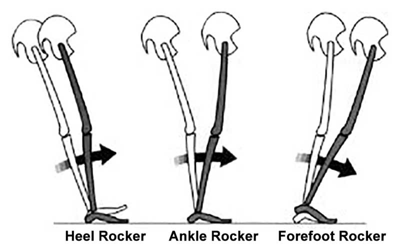
Geee....Looks like forefoot rocker really IS IMPORTANT, eh?

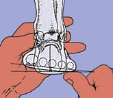
...and what have we been saying about being able to dorsiflex your big toe? Watch how well you or your client can descend the 1st ray (remember that if the head of the 1st does not go down and anchor, its axis of rotation moves dorsally and posterior, limiting dorsiflexion of the joint)

This article should make you look at the "toe break" in a shoe...

"They found that increasing bending stiffness assisted with propulsion during running, reducing the metabolic cost of running by about 1%. However, at a certain level, the increased elasticity began to interfere with the natural flexion of the first MTP joint, reducing the contribution of ankle joint torque to push-off and counteracting the metabolic benefits. Within the study population, the threshold of bending stiffness for optimal energetics varied significantly from one runner to the next, suggesting running shoe design may need to be tuned to an individual runner’s needs." 

http://lermagazine.com/news/in-the-moment-sports-medicine/stiffer-shoes-improve-running-energetics-as-long-as-first-mtp-flexion-is-preserved

tumblr_o80wf8tU9u1qhko2so1_400.jpg
tumblr_o80wf8tU9u1qhko2so2_1280.jpg

Do you know where your rocker is?

At 1st pass, some articles may seem like a sleeper, but there can be some great clinical pearls to be had. I recently ran across one of these. It was a presentation from the  42nd annual American Academy of Orthotists and Prosthetists meeting in Orlando, March 2016 entitled “ Shifting Position of Shoe Heel Rocker Affects Ankle Mechanics During Gait”. The title caught my eye.

They looked at ankle kinematics while keeping the toe portion of rocker constant at 63% of foot length, angled at 25 degrees and shifting the base of a rockered shoe from 1cm behind the medial malleolus, directly under it and 1cm anterior to it. Knee and hip kinematics did not differ significantly, however ankle range of motion did.

The more forward the ankle rocker, the less plantarflexion but more ankle dorsiflexion at midstance. So, the question begs, why do we care? Lets explore that further…

  • Think about the “average” heel rocker in a shoe. It largely has to do with the length of the heel and heel flare (base) of the shoe. The further back this is (ie; the more “flare”) the more plantar flexion at heel strike and less ankle dorsiflexion (and thus ankle rocker, as described HERE) you will see. Since loss of ankle dorsiflexion (ie: rocker) usually means a loss of hip extension (since these 2 things should be relatively equal during gait (see here), and that combination can be responsible for a whole host of problems that we talk about here on the blog all the time. Picking a shoe with a heel rocker based further forward (having less of a flare) would stand to promote more ankle dorsiflexion.
  • Having a shoe with a greater amount of “drop” from heel to toe (ie: ramp delta) is going to have the same effect. It will move the calcaneus forward with respect to the heel of the shoe and effectively move the rocker posteriorly.
  • Lastly, look a the shape of the outsole of the shoe. The toe drop is usually clear to see, but does it have a heel rocker (see the picture above)?

These are  a few examples of what to look for in a clients shoe when examining theirs or making a recommendation, depending on whether you are trying to improve or decrease ankle rocker. We can’t think of why you would want to decrease ankle rocker, but with conditions like rigid hallux limitus, where the person has limited or no dorsiflexion of the great toe, you may want to employ a rockered sole shoe. We would recommend one with the rocker set more forward.

Forefoot valgus: A fixed structural defect in which the plantar aspect of the forefoot is everted on the frontal plane relative to the plantar aspect of the rearfoot; the calcaneum is vertical, the mid tarsal joints are locked and fully pronatedWant…

Forefoot valgus: A fixed structural defect in which the plantar aspect of the forefoot is everted on the frontal plane relative to the plantar aspect of the rearfoot; the calcaneum is vertical, the mid tarsal joints are locked and fully pronated

Want to know more? Join us Wednesday evening: 5 PST, 6 MST, 7 CST, 8 EST for Biomechanics 309: Focus on the forefoot on onlinece.com.

McGraw-Hill Concise Dictionary of Modern Medicine. © 2002 by The McGraw-Hill Companies, Inc.

Fore foot types: Differences between forefoot varus and forefoot supinatus.Certainly this can be a contraversial topic. Perhaps this will help clear up some questions.Supination of the forefoot that develops with adult acquired flatfoot is defined a…

Fore foot types: Differences between forefoot varus and forefoot supinatus.

Certainly this can be a contraversial topic. Perhaps this will help clear up some questions.

Supination of the forefoot that develops with adult acquired flatfoot is defined as forefoot supinatus. This deformity is an acquired soft tissue adaptation in which the forefoot is inverted on the rearfoot. Forefoot supinatus is a reducible deformity. Forefoot supinatus can mimic, and often be mistaken for, a forefoot varus. A forefoot varus differs from forefoot supinatus in that a forefoot varus is a congenital osseous deformity that induces subtalar joint pronation, whereas forefoot supinatus is acquired and develops because of subtalar joint pronation (1).

A Forefoot Varus induces STJ pronation whereas a Forefoot Supinatus is created because of STJ pronation (2).

As the foot experiences increased subtalar joint (STJ) pronation moments during weightbearing activities (as in forefoot supinatus) , the medial metatarsal rays will be subjected to increased dorsiflexion moments and the lateral metatarsal rays will be subjected to decreased dorsiflexion moments. Over time, this increase in STJ pronation moments will tend to cause a lengthening of the plantar ligaments and medial fibers of the central component of the plantar aponeurosis and a shortening of the dorsal ligaments in the medial longitudinal arch. As a result, the influence of increased STJ pronation moments occurring over time during weightbearing activities will tend to cause the following (3):

1. An increase in inverted forefoot deformity.
2. A decrease in everted forefoot deformity.
3. A change in everted forefoot deformity to either a perpendicular forefoot to rearfoot relationship or to an inverted forefoot deformity.

More on the forefoot tomorrow evening on onlinece.com: Biomechanics 309. Join us!

1. Clin Podiatr Med Surg. 2014 Jul;31(3):405-13. doi: 10.1016/j.cpm.2014.03.009. Forefoot supinatus. Evans EL1, Catanzariti AR2.

2. https://kenva.wordpress.com/…/…/forefoot-varus-or-supinatus/

3. http://www.podiatry-arena.com/podiatry-forum/showthread.php…

Forefoot Varus or Forefoot Supinatus?Forefoot varus is a fixed, frontal plane deformity where the forefoot is inverted with respect to the rearfoot. Forefoot varus is normal in early childhood, but should not persist past 6 years of age (i.e. when d…

Forefoot Varus or Forefoot Supinatus?

Forefoot varus is a fixed, frontal plane deformity where the forefoot is inverted with respect to the rearfoot. Forefoot varus is normal in early childhood, but should not persist past 6 years of age (i.e. when developmental valgus rotation of forefoot on rearfoot is complete, and plantar aspects of fore- and rearfoot become parallel to, and on same plane as, one another (1)

Forefoot supinatus is the supination of the forefoot that develops with adult acquired flatfoot deformity. This is an acquired soft tissue adaptation in which the forefoot is inverted on the rearfoot. Forefoot supinatus is a reducible deformity. Forefoot supinatus can mimic, and often be mistaken for, a forefoot varus. (2)

A forefoot varus differs from forefoot supinatus in that a forefoot varus is a congenital osseous where a forefoot supinatus is acquired and develops because of subtalar joint pronation.

“Interestingly, only internal rotation of the hip was increased in subjects with FV – no differences were present in hip adduction and knee abduction between subjects with and without FV. The authors nevertheless conclude that FV causes significant changes in mechanics of proximal segments in the lower extremity and speculate that during high-speed weight-bearing tasks such as running, the effects of FV on proximal segments in the kinetic chain might be more pronounced.”

We wonder if the folks in this study had a true forefoot varus, or actually a forefoot supinatus (3).


The Gait Guys


1. Illustrated Dictionary of Podiatry and Foot Science by Jean Mooney © 2009 Elsevier Limited.

2. Evans EL1, Catanzariti AR2. Forefoot supinatus.
Clin Podiatr Med Surg. 2014 Jul;31(3):405-13. doi: 10.1016/j.cpm.2014.03.009.

3. Scattone Silva R1, Maciel CD2, Serrão FV3. The effects of forefoot varus on hip and knee kinematics during single-leg squat. Man Ther. 2015 Feb;20(1):79-83. doi: 10.1016/j.math.2014.07.001. Epub 2014 Jul 12.

This is a follow up to our last post on forefoot varus, available here.

Remember, ou are looking at a person with an uncompensated, rigid fore foot varus. This individual is not able to get the head of the 1st ray to the ground at all, and he has a Morton’s foot to boot (no pun intended). 
So, what do we see?

  • 1st of all, you will note his 2nd metatarsal is longer than his 1st. When he goes up on his toes, you see his foot invert and will see curling of the toes 3-5 in an attempt to stabilize the foot. 
  • You will also see his foot looks pretty flat. He has an arch (you can see it as he goes up onto his toes) and the “flatness” is actually due to the fore foot varus.
  • You will see a bunion forming bilaterally, due again to the uncompensated fore foot varus, and his inability to anchor the head of the 1st metatarsal. 
  • The posterior view shows relatively vertical calcaneii (no no rearfoot valgus), but you can really see the effects of the fore foot varus, with medial fall of the midfoot.
  • note the prominent “pump bumps” on the lateral calcaneus biaterally, from chronic rubbing on the shoes. 
The Gait Guys. Getting you closer to being a foot nerd with each post.
 
Lost? Having trouble with all these terms and nomenclature? Take our national shoe fit program, available by clicking here.

The Gait Guys. Uber foot geeks. Still bald and good looking. Separating the wheat from the chaff, with each and every post.

tumblr_ms7kvihT0Y1qhko2so1_1280.jpg
tumblr_ms7kvihT0Y1qhko2so2_1280.jpg

What do we have here and what type of shoe would be appropriate?

You are looking at a person with a fore foot varus. This means that the fore foot (ie, plane of the metatarsal heads) is inverted with respect to the rear foot (ie, the calcaneus withe the subtalar joint in neutral). Functionally translated, this means that they will have difficulties stabilizing the medial tripod (1st MET head) to the ground making the forefoot and arch unstable and likely rendering the rate and degree of pronation increased.

Having trouble with terminology? check out this post on FF varus.

The incidence of this condition is 8% of 116 female subjects (McPoil et al, 1988) and 86% of 120 male and female subjects (Garbalosa et al, 1994), so it happens more in males.

Fore foot varus occurs in 3 flavors:
  • compensated
  • uncompensated
  • partially compensated
What is meant by compensated, is that the individual is able to get the head of the 1st ray to the ground completely (compensated), partially, or, when not at all, uncompensated.
What this means from a gait perspective ( for partially and uncompensated conditions) is that the person will pronate through the fore foot to get the head of the 1st ray down and make the medial tripod of the foot (ie, they pronate through the subtalar joint to allow the 1st metatarsal to contact the ground). This causes the time from mid-stance to terminal stance to lengthen and will inhibit resupination of the foot. We will have an upcoming additional post on this soon and will put a link here when we do.
Today we are looking at a rigid, uncompensated forefoot varus, most likely from insufficient talar head derotation during fetal development and subsequent post natal development. They will not get to an effective foot tripod. They will collapse the whole foot medially. These people look like severely flat-footed hyperpronators.

So, what do you do and what type of shoe is appropriate? Here’s what we did:
  • try and get the 1st ray to descend as much as possible with exercises for the extensor hallucis brevis and short flexors of the toes (see our videos on youtube)
  • create more motion in the foot with maniipuulation, massage mobilization to optimize what is available
  • strengthen the intrinsic muscles of the feet (particularly the interossei
  • increase strength of the gluteus maximus and posterio fibers of the gluteus medius to slow internal rotation of the leg during initial contact to midstance
  • put him in a flexible shoe for the 1st part of the day, to exercise the feet and a more supportive, medially posted (ideally fore foot posted) shoe for the latter part of the day as the foot fatigues
  • monitor his progress at 3-6 month intervals
  • a rigid orthotic will likely not help this client and they will find it terribly uncomfortable because this is a RIGID deformity for the most part (the foot will not accommodate well to a corrective orthotic. Besides, the correction really has to be made at the forefoot anyways. We will talk about medial forefoot postings again at a later date.)
Lost? Having trouble with all these terms and nomenclature? Take our national shoe fit program, available by clicking here.

The Gait Guys. Uber foot geeks. Still bald and good looking. Separating the wheat from the chaff, with each and every post.

A Tale of Two Footies
Time for a pedograph, folks. What do we have here? Look at the last analysis here. 
To review :
Let’s divide the foot into 3 sections: the rear foot, the mid foot and the fore foot.
First of all, are they symmetrical? Look care…

A Tale of Two Footies

Time for a pedograph, folks. What do we have here? Look at the last analysis here.

To review :

Let’s divide the foot into 3 sections: the rear foot, the mid foot and the fore foot.

First of all, are they symmetrical? Look carefully at the fore foot on each side. NO! the right foot looks different than the left, so we are looking at asymmetrical pathology.

Let’s start at the rear foot: The heel teardrop is elongated on both sides, slightly more on the right; this means incraesed calcaneal eversion (or rearfoot pronation) bilaterally, R > L. The right heel shows increased pressure (more ink = more pressure).

Next up, the mid foot. Similar shapes, more pressure and printing on the left. Did you notice the “tail” of the 5th metatarsal printing, giving it a wider print? This person is staying on the outside of their foot longer than normal, right (more ink) more than left.

How about the fore foot? Lots going on there.

Lets start on the left

Notice the mild increased printing of the 5th and 4th metatarsal heads. Force should be traveling from lateral to medial here, as the foot goes into supination. A relatively normal amount of pressure on the head of the 1st metatarsal.

Now look at the toes. Notice that space between the 2nd and 3rd? This gal had an old fracture and has an increased space between them.

Now how about the right?

Increased pressure on most of the heads with a concentration on the 1st metatarsal. Hmmm…what would cause that? this is typical of someone who has a 1st ray (cunieform and metatarsal) that is hypomobile, such as with someone with a forefoot valgus (as this person does) or a dropped 1st metatarsal head (which is usually rigid, as is NOT the case here).

Did you see that rpinting at the medial aspect of the proximal phalanyx of the hallux (ie. big toe)? This gal externally rotates the lower extremity to push off the big toe to propel herself forward. This is because the 1st metatarsal head hits the ground BEFORE the 5th (as we would normally expect to see, like in the left foot), and because the weight is now on the outside of the foot, she need to push off SOMETHING.

Getting better at this? We hope so. Keep reading the blog and look at some of our past pedograph posts here.

The Gait Guys. Teaching you about the importance of gait, each and every day!