

It makes sense…but which came 1st?
Just make sure you ask your foot patients about their back, and your back patients about their feet
The Gait Guys
It makes sense…but which came 1st?
Just make sure you ask your foot patients about their back, and your back patients about their feet
The Gait Guys


Subtle clues. Helping someone around their anatomy
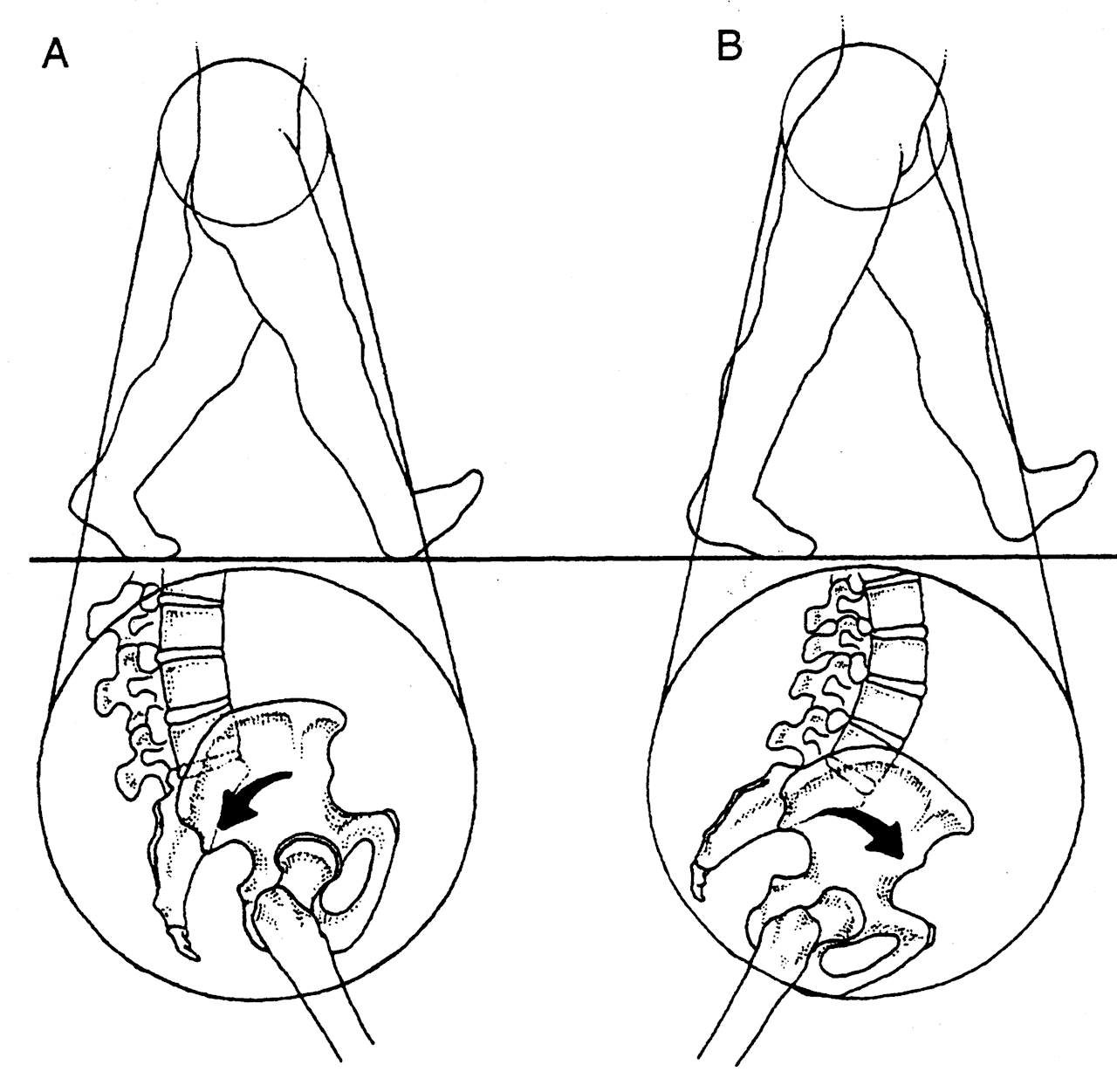
This patient comes in with low back pain of years duration, helped temporarily with manipulation and activity. Her exam is relatively benign, save for increased lumbar discomfort with axial compression in extension and extension combined with lateral bending. Believe it or not, her abdominal and gluteal muscles (yes, all of them) test strong (no, we couldn’t believe it either; she is extremely regular with her exercises). She has bilateral internal tibial torsion (ITT) and bilateral femoral retro torsion (FRT). She has a decreased progression angle of the feet during walking and the knees do not progress past midlilne. There is a loss of active ankle rocker with gait, but not on the exam table; same with hip extension.
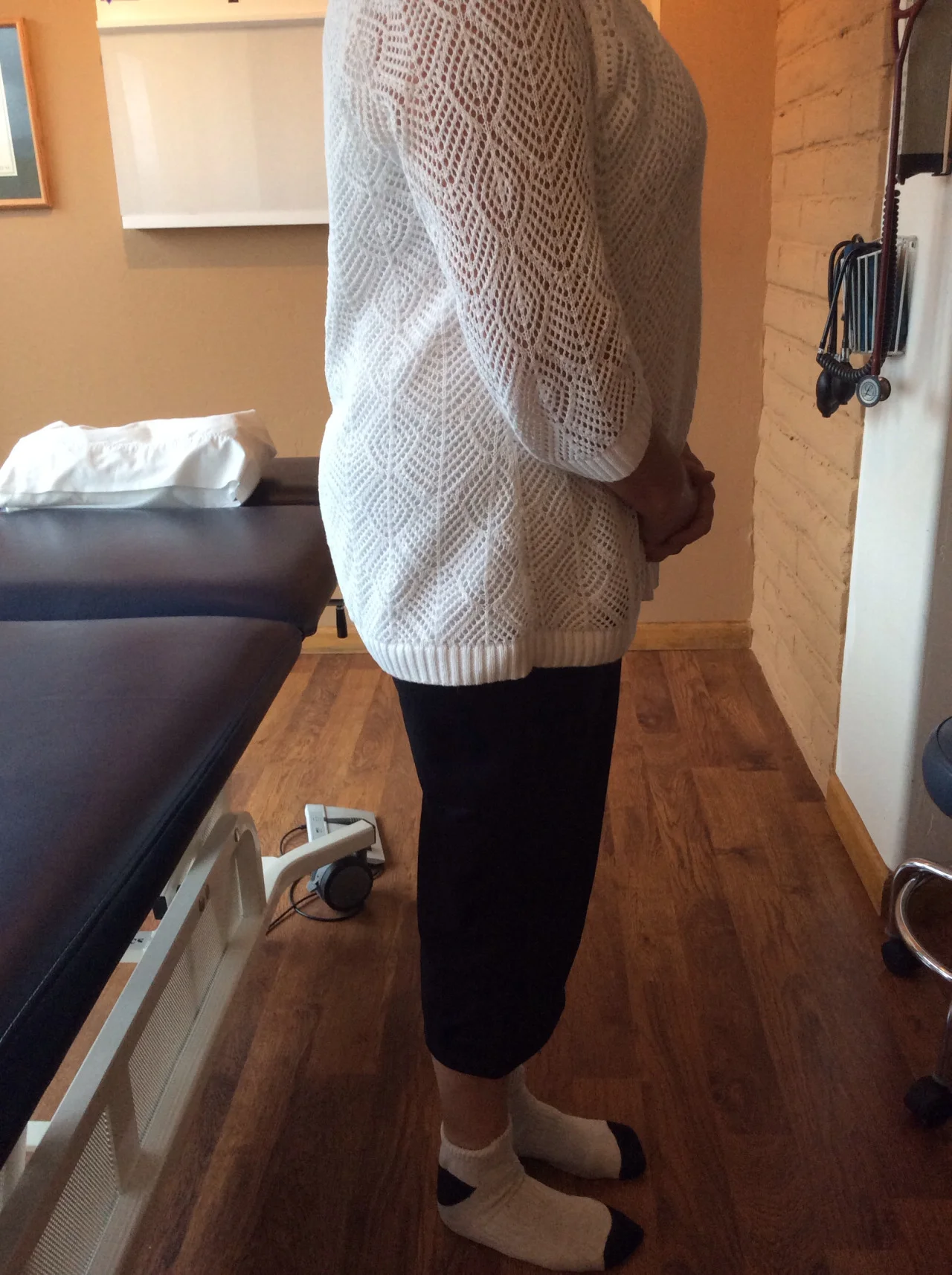
We know she has a sweater on which obscures things a bit, but this is what you have to work with. Look carefully at her posture from the side. The gravitational line should pass from the earlobe, through the shoulder, greater trochanter and through or just anterior to the lateral malleolus.
In the top picture, can you see how her pelvis is anterior to this line? Do you see how it gets worse when she lifts her hands over her head (yes, they are directly over head)? This can signify many things, but often indicates a lack of flexibility in the lumbar lordosis; in this case, she cannot extend her lumbar spine further so she translates her pelvis forward. Most folks should have enough range of motion from a neutral pelvis and enough stability to allow the movement to occur without a significant change. Go ahead, we know you are curious, go watch yourself do this in a mirror and see if YOU change.
Looking at the bottom left picture, can you pick out that she has a genu valgus? Look at the hips and look at the tibial angle.
In the bottom left picture, did you note the progression angle (or lack of) in her feet? This is a common finding (but NOT pathognomonic) in patients with internal tibial torsion. Notice the forefoot adductus on the right foot?
So what do we think is going on?
What did we do?
why didn’t we put her in an orthotic to externally rotate her lower extremity? Because with internal tibial torsion, this would move her knee outside the saggital plane and create a biomechanical conflict at the knee and possibly compromising her meniscus.
Cool case, eh? We thought so. Keep on learning so your brain keeps expanding. If you are not growing your brain, you are shrinking it!
The Gait Guys

Donate here


Help keep us free ! Make a donation by clicking. here.

Donate to get exclusive content from The Gait Guys
OUR SEARCH BOX IS INTUITIVE, TYPE IN YOUR KEY WORD, WAIT, THEN SCROLL DOWN.
Email us: our email is found under the "Disclaimer" Tab above.

Powered by Squarespace.