tumblr_nn1ca5fx0C1qhko2so1_r1_1280.jpg
tumblr_nn1ca5fx0C1qhko2so2_r1_1280.jpg
tumblr_nn1ca5fx0C1qhko2so3_r1_1280.jpg
tumblr_nn1ca5fx0C1qhko2so4_r1_1280.jpg

Simple Foot Exercises are effective!

Conclusion “These results suggest that the toe spread out (TSO) exercise can be recommended for preventing or correcting HV deformity at an early stage.”

We know and teach that foot exercises work. Here is a nice objective paper (click underlined for abstract) on two exercises we prescribe often.

Here is our variation of the TSO exercise we call the “Lift, Spread and Reach” exercise

Stand comfortably with your feet about shoulder width apart

Stand on your foot tripod with your toes extended. Concentrate on feeling pressure at the center of the calcaneus, the head of the 1st metatarsal and the head of the 5th metatarsal

Lift your toes as high as possible

Spread out (abduct) your toes as much as possible

Reach forward with your toes as far as possible

Place your toes back don on the ground as flat as possible.

repeat 10 X

You can augment the exercise with a rubber band around the toes to provide resistance after you can perform the exercise competently.

Happy exercising!

Ivo and Shawn


Kim MH1, Kwon OY, Kim SH, Jung DY.: Comparison of muscle activities of abductor hallucis and adductor hallucis between the short foot and toe-spread-out exercises in subjects with mild hallux valgus

J Back Musculoskelet Rehabil. 2013;26(2):163-8. doi: 10.3233/BMR-2012-00363.

tumblr_nhdevsLYyN1qhko2so3_400.jpg
tumblr_nhdevsLYyN1qhko2so4_r1_250.jpg
tumblr_nhdevsLYyN1qhko2so1_1280.jpg
tumblr_nhdevsLYyN1qhko2so2_250.jpg

Keeping it Objective.

For clinicians and some die hard foot geeks, we often like to keep things objective. What could be more objective than an angular measurement? A few important measurements when examining or radiographing feet can give us information about clinical decision making (not that we suggest radiographs for mensuration purposes unless you are a surgeon, but when they are already available, why not put them to good use ?). When things fall outside the accepted range, or appear to be heading that way, these numbers can help guide us when to intervene. 

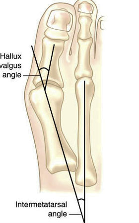
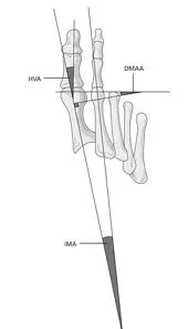
Hallux valgus refers to the big toe headed west (or east, depending on the foot and your GPS). In other words, the proximal and distal phalanyx of the great toe (hallux) have an angle with the 1st metatarsal shaft of typically > 15 degrees. This angle, called the Hallux Valgus Angle (HVA above) is used to judge severity, often for surgical intervention purposes but can guide conservative management as well. 

Metatarsus Primus Varus (literally, varus deformity of the 1st metatarsal) often accompanies Hallux Valgus. It describes medial deviation of the 1st metatarsal shaft, greater than 9 degrees. This angle is called the intermetatarsal angle and is measured by the angle formed by lines drawn parallel along the long axis of the 1st and 2nd metatarsal shafts. 

One other measurement is the Distal Metatarsal Articular Angle, which measures the angle between the metatarsal shaft and the base of the distal articular cap (ie, where the cartilage is) of the 1st metatarsal. This typically should be less than 10 degrees, preferably less than 6 degrees. Remember, these are static angles, things can change with movement, engagement, weight bearing strategies and shoes. What you see statically does not always predict dynamic angles and joint relationship.s

Are you doing surgery? Perhaps, as a last resort. Hallux valgus and metatarsus primus varus can be treated conservatively.

How do you do that?

The answer is both simple and complex.

The simple answer is: anchor the head of the 1st ray and normalize foot function. This could be accomplished by:

  • EHB exercises to descend the head of the 1st metatarsal
  • exercise the peroneus longus, to assist in descending the head of the 1st metatarsal
  • short flexor exercises, such as toe waving, to raise the heads of the lesser metatarsals relative to the 1st
  • work the long extensors, particularly of the lesser metatarsals to create balance between the flexors and extensors
  • consider using a product like “Correct Toes” to normalize the pull of the muscles and physically move the proximal and distal phalanyx of the hallux
  • wear shoes with wide toe boxes, to allow the foot to physically splay
  • consider using an orthotic with a 1st ray cut out, to help descend the head of the 1st metatarsal

This is by no means an exhaustive list and you probably have some ideas of your own. 

The complex answer is that in the above example, we have only included conservative interventions for the foot and have not moved further up the kinetic (or neurological chain). Could improving ankle rocker help create more normal mechanics? Would you accomplish this by working the anterior leg muscles, the hip extensors, or both? Could a weak abdominal external oblique be contributing? How about a faulty activation pattern of the gluteus medius? Could a congenital defect or genetic be playing a role? We have not asked “What caused this to occur in the 1st place?”

Examine your patients and clients. Understand the biomechanics of what is happening. Design a rehab program based on your findings. Try new ideas and therapies. it is only through our failures that we can truly learn.

The Gait Guys

references used:

http://www.bjjprocs.boneandjoint.org.uk/content/90-B/SUPP_II/228.3

http://www.slideshare.net/ANALISIS/hallux-valgus-2008-pp-tshare

http://www.orthobullets.com/foot-and-ankle/7008/hallux-valgus

http://www.slideshare.net/bahetisidharth/hallux-valgus-31768699?related=1

tumblr_nhbuqbFrTD1qhko2so1_250.png
tumblr_nhbuqbFrTD1qhko2so2_500.png
tumblr_nhbuqbFrTD1qhko2so3_r1_1280.jpg

When the big guy heads medially….Game Changer

Lately we have been seeing a lot of bunions (hallux valgus). While doing some research on intermetatarsal angles (that’s for another post) we came across the nifty diagram you see above. 

Regardless of the cause, as the 1st metatarsal moves medially, there are biomechanical consequences. Lets look at each in turn. 

  • the EHB (extensor hallucis brevis) axis shifts medially. this muscle, normally an extensor of the proximal phalanyx, now becomes more of an abductor of the hallux. It’s secondary action of assisting the descent of the head of the 1st metatarsal no longer happens and it actually moves the base of the proximal phalanyx posteriorly, altering the axis of centration of the joint, contributing to a lack of dorsiflexion of the joint and a hallux limitus
  • Abductor hallucis becomes more of a flexor, as it moves to the plantar surface of the foot. Remember, a large percentage of people already have this muscle inserting more on the plantar surface of the foot (along with the medial aspect of the flexor hallucis brevis), so in these folks, it moves even more laterally, distorting the proximal phalanx along its long axis (ie medially) see this post here for more info
  • Flexor hallucis brevis moves more laterally. Remember this muscle houses the sesamoid bones before inserting onto the base of the proximal phalannx; the medial blending with the abductor hallucis and the lateral with the adductor hallucis. Because the sesamoid bones have moved laterally, they no longer afford this muscle the mechanical advantage they did previously and the axis of motion of the 1st metatarsal phalangeal joint moves dorsally and posterior, contributing to limited dorsiflexion of that joint and a resultant hallux limitis. The lateral movement of the sesamoids also tips the long axis of the 1st metatarsal and proximal phalanyx into eversion. In addition, the metatarsal head is exposed and is subject to the ground reactive forces normally tranmittted through the sesamoids; often leading to metatarsalgia. 
  • Adductor hallucis: this muscle now has a greater mechanical advantage  and because the head of the 1st ray is not anchored, acts to abduct the hallux to a greater degree. The now everted position of the hallux contributes to this as well

As you can see, there is more to the whole than the sum of the parts. Bunions have many biomechanical consequences, and these are only a small part of the big picture. Take you time, learn your anatomy and examine everything that has a foot!

See you in the shoe isle…

Ivo and Shawn

pictures from: http://www.orthobullets.com/foot-and-ankle/7008/hallux-valgus and http://www.stepbystepfootcare.com/faqs/nakedfeet/