tumblr_mg6a1wNO4i1qhko2so1_1280.jpg
tumblr_mg6a1wNO4i1qhko2so2_400.jpg

A little neuro, anyone?

Welcome to Monday, and yes, it is a NEURO day. In fact, if you got up this morning, you too are having a NEURO day. Dr Allen thinks it’s all about the ORTHOPEDICS, but without NEURO, there would not be any orthopedics : )

A dialogue from one of our avid readers, Dr. Ryan.

Dr. Ryan: Hey Ivo,

I just read this article on Mercola’s site which is an interview with Dr. Craig Buhler who does muscle activation techniques.  Can you check this for accuracy?  This must be a mistake b/c I always thought spindle activation will facilitate the muscle to contract.  Also, I always wondered why the O/I attachment points are tender in muscles that are inhibited.  Does his description sound right to you.  If not, do you have a better explanation?

“Your muscle system and nervous system relate to each other from within tiny muscle fibers called spindle cells, which monitor stretch. If your muscle is overloaded too rapidly, the spindle cells will temporarily inhibit the muscle. The next time you contract the muscle, it will fire again. Similarly, cells within your tendons called Golgi tendon organs also measure and monitor stretch. If your tendon is stretched too rapidly or exceeds its integrity, the Golgi tendon organs will temporarily inhibit the muscle. But the next time the muscle fires, it will again fire appropriately.

"But there’s a fail-safe system," Dr. Buhler explains. "It’s where the tendon attaches into the periosteum of the bone and the little fibers there are called Sharpey’s fibers. Those fibers are loaded with little receptors that monitor tension. And if the integrity of those fibers are exceeded, they inhibit the muscle, just like a circuit breaker would inhibit an electrical circuit.

Once that happens, the muscle will still fire under passive range of motion. But if you load the muscle, it gives way. If you continue to load the muscle, your body creates pain at the attachment points to protect you. What the central nervous system does at that point is compute an adaptive strategy by throwing stress into the muscle next to it. Other tissues begin to take on more of the load for the muscle that’s been injured.”

Here is a link to the entire article if you want to check it out:

http://fitness.mercola.com/sites/fitness/archive/2013/01/04/advanced-muscle-integration-technique.aspx?e_cid=20130104_DNL_art_1"

Dr Ivo: Thanks Dr. Ryan.

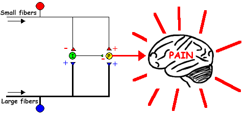
Spindles monitor length and GTO’s monitor tension. My understanding is spindles, when activated, stimulate the alpha motor neuron(at the cord) and cause contraction of that muscle or motor unit. GTO’s, when activated, cause inhibition of the muscle they are associated with. I am not aware of them being inhibitory, only GTO’s. They are believed to be GABAnergic synapses. The impulse (at least in cats) can be smaller or inhibited if the muscle is held in contraction for an extended period of time (see attached)

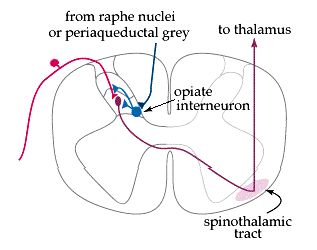
Perhaps he is talking about spindle dysfunction, where the intrafusal portion of the spindle (which is innervated by a gamma motor neuron) is either excited or inhibited. The gamma’s are more of a slave to the interneuronal pool (in the cord), which would be the sum total of all excitatory and inhibitory input to that area (ie the central integrated state). This not only reflects local receptor input but also cortical information descending (from areas 4s and 6 in the precentral gyrus) AND descending information from the caudal reticular formation.

Based on what you sent, I do not agree with the 1st 2 sentences. I was not aware about increased receptor density of Sharpeys fibers. I did a quick search and found this: http://www.ncbi.nlm.nih.gov/pmc/articles/PMC2100202/  , which eludes to it and here: http://www.ncbi.nlm.nih.gov/pmc/articles/PMC3098959/. I will have to dive in more when I have time.

Not sure why O/I attachments are tender in inhibited muscles. I find them tender in most folks. Maybe because inhibited muscles ave altered receptor function and that preloads the nociceptive afferent pathway or at least that neuronal pool? Are they closer to threshold for some reason? Not sure. LMK what you find.

Thanks for getting me jazzed about sharpeys fibers!

for those of you who need to know YES, there will be a forthcoming Sharpeys fibers article

Dr. Ryan: That’s what I thought.  Thanks for looking into it and I will check out those links.  You have jazzed me plenty of times over the years.  Glad I could jazz you up for a change.  Have a great weekend.


Yes, Dr Ivo is definitely an uber neuro geek, especially when he spends time on the weekend talking about spindles!


all material copyright 2013 The Homunculus Group/ The Gait Guys. All rights reserved. Please as before you lift our stuff.

Neuromechanics?  This early in the morning?
It has been a while since we have done a neuromechanics post. While doing some research for one of our PODcasts, We ran across this paper: http://www.ajronline.org/content/184/3/953.full
It’s title?
…

Neuromechanics?  This early in the morning?

It has been a while since we have done a neuromechanics post. While doing some research for one of our PODcasts, We ran across this paper: http://www.ajronline.org/content/184/3/953.full

It’s title?

Midbrain Ataxia: An Introduction to the Mesencephalic Locomotor Region and the Pedunculopontine Nucleus

Yikes! What a mouthful!

What’s the bottom line?

The paper review a condition called “gait ataxia”. In plain English this means “aberrant or unsteady” gait. Things which usually cause gait ataxia originate in an area of the brain called the cerebellum, which coordinates all muscle activity. If you drink to much alcohol, it affects your cerebellum and you have a “wobbly” gait : ).

This paper looks at another area of the brain called the midbrain. It is the top part of the brainstem and contains an important gait integration and initiation center called the “midbrain locomotor nucleus”. The paper looks at 3 different cases and has some cool MRI images to see, along with alot of fancy neurological words and pathways.

Whenever we see gait ataxia, we think of impaired proprioception (look here for a bunch of posts on that, or at this post specifically).

There are many factors to consider when evaluating ataxic (or wobbly) gait, and this just gives us all one more place to look.

The Gait Guys. Making you smarter every day!

tumblr_m69aylFcmg1qhko2so1_500.gif
tumblr_m69aylFcmg1qhko2so2_500.gif

Neuromechanics Weekly: How does appropriate movement diminish pain?

We talk about proper (or should we say appropriate) movement (including gait) inhibiting or diminishing pain. So, how does that happen?

Above on the left is a great diagram that we will work through.

You are looking at a cross section of a spinal cord (We can hear the groans already!) We remember that the dorsal horn (posterior part) is sensory and the ventral horn (front) is motor. In between them (the lateral horn) is autonomic (this runs automatized body functions such as your heart, lung, guts, etc).

Small nerve fibers subserve pain. These are the A delta and C nerve fibers. “Small” refers to fiber diameter of the nerve. These nerves are where pain stimuli enters the spinal cord; they enter the sensory dorsal horn and synapse/connect there with the next neuron in line that takes the sensory message up the spinal cord to the brain to tell you about the pain including its intensity, location etc. Pain can result from tissue damage or injury (which can be due to, or the result of, poor biomechanics).

Large diameter nerve fibers subserve sensations like touch, pressure, vibration, muscle spindles (muscle length) and golgi tendon organs (muscle tension/load). These fibers also enter the sensory dorsal horn, but they do not synapse immediately, unlike pain fibers. They ultimately travel up to the top of the brainstem or cerebellum to coordinate information with other data your brain is processing. They send a branch (or collateral) to an inhibitory neuron, which excites the inhibitory neuron. Thus, if you excite an inhibitory neuron, it does it’s job and inhibits the propagation of an impulse. In this case, it inhibits the pain impulse from traveling to the cortex. So pain is inhibited. Appropriate biomechanics excite the largest population of receptors and provide the most effective response. 

Now look at the diagram on the right. It is a simplified schematic of the one on the left, with detail of the connections. Note that the LARGE FIBERS (from joint mechanoreceptors, spindles, muscles, etc) EXCITE the inhibitory interneuron (which would inhibit it). Also note that the SMALL FIBERS INHIBIT the inhibitory internuron (which would excite it!)

There you have it. Clear as mud? Go through some of our old posts on receptors and FEEL THE PAIN (parts 1, 2, 3+4) and come back to this and read it again. You know you want to be a geek, so go ahead!

The Gait Guys: Geeks on many levels. helping you to presynaptically inhibit pain on a daily basis, through better movement.

Neuromechanics on Saturday?
We have long been talking about the importance of the cerebellum in gait and motor activity (see here).
Here is a study (Non-Invasive Brain Stimulation Shown to Impact Walking Patterns) that looks at a new technique for u…

Neuromechanics on Saturday?

We have long been talking about the importance of the cerebellum in gait and motor activity (see here).

Here is a study (Non-Invasive Brain Stimulation Shown to Impact Walking Patterns) that looks at a new technique for using electrical stimulation of the brain’s cerebellum (trans cranial direct current stimulation to be exact) to change gait on a split belt treadmill (a double treadmill where each leg moves a slightly different speed). The study found that during the electrical stimulation the anode (negative charge) seems to speed up the learning process (our theory: more electrons, possibly creating a temporary electrical gradient which depolarizes (excites) the cells to a greater degree). And the cathode seemed to slow things down (our theory, it hyperpolarizes the cell and makes it less excitable).

Take home message? There are new neurologic studies and experiments that may be proving helpful in retraining gait function.  Stimulating the brain’s cerebellum seems to speed up learning or slow it down, depending on your client’s needs. We are sure we will be seeing more of this kind of stuff at technology advances. 

Maybe Larry Niven wasn’t that far off. (We loved the story “The Long Arm of Gil Hamilton”).  This could be a great, non invasive tool for rehab (or maybe improving performance!)

The Gait Guys…taking you deeper down the rabbit hole…

Neuromechanics Weekly:
Gait NoiseThink of “Gait Noise” as those things which alter the sum total of all neuronal activity acting at a specific locus. Gait noise is all of the aberrant signals that distort the correct and most functionally desirable …

Neuromechanics Weekly:

Gait Noise

Think of “Gait Noise” as those things which alter the sum total of all neuronal activity acting at a specific locus. Gait noise is all of the aberrant signals that distort the correct and most functionally desirable signal necessary for a clean gait.  Think of noise as the static found between radio stations, that irritating white noise that blurs out the perfect radio station from coming in clearly.  Gait noise is thus anything that impairs a clear sensory and motor signal to and from the central nervous system required for clean uncompensated gait. These definitions will help you understand where we are going with this.

1. Communication: Anything that interferes with, slows down, or reduces the clarity or accuracy of a communication. Thus, superfluous data or words in a message are noise because they detract from its meaning.
2. Quality control: Variability that may be caused by changes in the ambient conditions, faulty machine performance, or uneven quality of the material or human factor inputs.
3. Telecommunications: Random disturbance introduced into a communication signal, caused by circuit components, electromagnetic interference, or weather conditions. Also called line noise.

Gait noise is therefore very undesirable. It could be interpreted as seeing a foot turned out more than normal, more than the other side. Seeing that compensation is a motor impairment and an undesirable motor pattern, but it also sends aberrant sensory information back into the nervous system. Bad information in, bad information out, and a viscous cycle ensues.  Gait noise can occur from a total knee replacement, from a scar, from a sprain, a broken bone, from the numbness of a diabetic neuropathy etc.  These all cause impairment of the sensory-motor-sensory loop.  Gait Noise theoretically could occur anywhere along the neuraxis (spinal cord and brain-brainstem) or even the peripheral nervous system, but it makes most sense to think of it happening where neurons congregate; most likely at synapses, especially at the spinal cord level. The wiring of the nervous system extends to all tissues, so the noise can occur anywhere for almost any reason.

In the words of Dr Ted Carrick, “Is the lesion at the receptor, the effector, the peripheral nerve, the spinal cord, the brainstem, the thalamus, the cerebellum or cerebrum?”  Where is the problem in otherwords ?  Lets explore how this relates to “Gait Noise”.

Today lets look at the receptor. Receptors are the information gatherers of the nervous system. Think of your 5 senses (vision, smell, taste, sound and touch). These are all subserved by receptors. Vision and touch seem to most affect gait and movement. This post will concentrate on touch.

Touch encompasses not only physical touch but also proprioception (see here for review of proprioception and receptors). These receptors: Pacinian corpuscles, Merkel discs, end bulbs of Krause, bare nerve endings, joint mechanoreceptors, muscle spindles and Golgi tendon organs are all included here. These sentinels provide the central nervous system (CNS) with mechanical environmental information, for comparison with that same information (there is much redundancy in the CNS) and other environmental information (balance, vision, hearing, etc), so that you can formulate (consciously or subconsciously) a response.

In short, there are multiple systems converging, in this case, on the peripheral receptor. Remember, receptors can be activated in may ways. A touch receptor could be activated not only by touch, but also by heat, cold, pressure, or even chemical (metabolic or toxic) means. Just like you may be a great tennis player, you could probably play racquet ball, handball, or football, even if you never played before. You may not excel, but you could get by. Receptors are no different; they may be BEST activated by touch, but other means could certainly do the job. This inadvertent activation creates receptor bias, as we like to call it “noise”, and that information is sent to the CNS for processing. If a touch receptor is activated, it is activated, and the CNS  sees it as an activation, whether it is intentional or not. These mixed signals are then processed along with everything else, creating “noise”.  And the noise might not be a desired signal. And these signals can be what initiates a gait change, a compensation, whether it be from information mis-processing or a strategy to cope.

Think of the application to your gait analysis, next time you are seeing something that you think you shouldn’t be seeing.  This is the problem with video gait analysis (as we take a moment to pound the wall on this topic ONE MORE TIME !).  What we see on a video analysis is not necessarily the problem, nor does what you see warrant a correction or a specific shoe. What we are seeing on video is their coping strategy after all of the CNS signals (noise and non-noise) have been processed, it is what they can do with what is available to them and with what makes most sense to the brain.  We have said before, as a classic example, that an over pronating foot might be a necessity to compensate for lack of internal hip rotation because the brain deems that functional pathology as more damaging at the hip than the hyperpronation at the foot.  Who are we to deem that the foot needs an orthotic or a stability shoe because of what we see?  Who are we to think that we can outsmart all the sensory-motor calculations of that persons brain without knowing all of the functional limitations of their body ? Perhaps if we take an hour to assess our client, and then see them for another visit or two, we can then correlate the gait video, our findings and our corrective work and then truly qualify a logical reasoning.  But this is a far more difficult game that this simple gait video or foot plantar pressure digital foot mapping nonsense.

Ivo and Shawn; the voices in your head, helping you sift out the noise.

tumblr_m2j60zuBR31qhko2so1_1280.jpg
tumblr_m2j60zuBR31qhko2so2_r2_1280.jpg

Neuromechanics Weekly: PART 2:

Stretching increases strength in contralateral muscles?

Lots of cool links in this post. please try and find time to check them out.

Figure it out?  Ever wonder about some of the magic behind some of those manual therapy techniques that are out there ? Sometimes it is not magic at all !

There are 2 related reasons we can think of to cause this seemingly odd length-strength phenomenon (OK, there are more, but this is what we are going to cover today):

  • Reciprocal Inhibition
  • Crossed extensor reflexes/responses

We remember reciprocal inhibition (as demonstrated in LEFT picture above) is when we activate or stimulate a muscle, the Ia afferent from that muscle stimulates that same muscle to contract (this is how a simple reflex arc works) and, through an inhibitory interneuron, inhibits the antagonist muscle on the opposite side of the joint.

Remember, that Ia afferents go to muscle spindles (don’t remember? look here); they respond to LENGTH changes. Wouldn’t you say stretching affects length? If we were talking about the R tricep surae group, we would be inhibiting the R anterior compartment.

But wait, the article said it affects the opposite side….Of course, there is more…

The picture on the right shows the crossed extensor response or reflex (don’t remember? look here). In a nutshell, when you FIRE the flexors on one side, you INHIBIT the extensors on the same side (sound like reciprocal inhibition? It should… it is : ) You also FIRE the extensors on the opposite side while INHIBITING the flexors on the opposite side. (Yes, the opposite side extensors will inhibit the opposite side flexors as well. Yes, this is also reciprocal inhibition).

But wait, that means the opposite calf would be weaker, not stronger, right?

It would be weaker if being called upon to be used at that moment in time, BUT in the study, stretching increased ROM of the stretched calf 8%, with a 1% loss of ROM of the opposite calf (study summary).

Hmm… sounds like shortening to me. That would mean that those spindles (ie the opposite calf)  would be MORE RESPONSIVE to stretch (ie a change in length; and coincidentally, the Golgi’s more responsive to the tension change) . And what happens when we preload a neuronal pool? The likelihood of firing is increased (like doing a Jendrassik maneuver to increase a reflex). The rest is neural adaptation (strength gains initially are due to increased efficiency of the nervous system. For a review to see our video on this, click here)

Interesting that one of the comments on the article was “I don’t have the full text of the paper but a summary prepared by Chris Beardsley and Bret Contreras states that one of the mechanisms for crossover in the case of unilateral strength training is thought to be modulation at the spinal cord level.”   Could they be talking about reciprocal inhibition and crossed extensor responses?

Wow! Very cool! And to think, you knew the answer. We are proud of you!

Ivo and Shawn…Neuro Geeks too!  And applying it to gait, running and motor patterns of all types !

tumblr_m2j5t6IDzZ1qhko2so1_1280.jpg
tumblr_m2j5t6IDzZ1qhko2so2_r1_400.gif

Neuromechanics Weekly: PART 1:

Stretching increases the strength in contralateral muscles?

The question is why (isn’t that always the question?). Applying your knowledge of neurology, can you figure it out?

Here’s a hint: There are at least 2 related reasons. See the pictures above and check back at noon for our theory.

Stew on this for an hour, seriously !   Engage your brain on this very important topic and we will see you in one hour.  For the next hour as you stretch, or stretch athletes, or patients or as you prescribe stretching as some homework to someone, think about how stretching one muscle creates strength in a contralateral muscles.  If you learn to use the power of the nervous system to your advantage you can get much better results.  The Gait Guys do.


Click above for the article

Neuromechanics Weekly: Gait and Running and the Crossed Extensor Responses

This week we discuss why upper and lower limbs are paired in gait, and the neuronal wiring that is involved.

Yup, this is pretty geeky stuff, but geez…isn’t it nice to know WHY something works? Think of the implications if YOU DON’T see the upper and lower limb pairing. Think of the implications during rehab. One limb SHOULD be doing the opposite of the other AND always look at the upper limbs and arm swing. Yes, the central  nervous system is involved. It is more than just biomechanics, perhaps this is why this stuff comes easier to us because of our deeper neurology background. The whole is greater than the sum of the parts….

Ivo and Shawn

Under Armor mouth Guards: Neuromechanics?

This week in neuromechanics weekly, we will look at the concept or preloading motor neuronal pools. A reader asked if we could look at the above link (click title) and offer some clinical commentary and some rather bold statements
Here are our thoughts:


The findings they speak of are not surprising at all…

The Temporomandibular joint (TMJ) is blessed with many mechanoreceptors and receives innervation from Cranial Nerve V (trigeminal nerve) and the upper cervical spine. There is physiological overlap through the trigeminocervical nucleus (in the upper midbrain or mesencephalon, the principal sensory nucleus) which receives the same innervation from the trigeminal nerve distribution and the upper 4 cervical neuromeres(nerve levels) (so double input into same pathway). Nicoli Bogduk published abody of research on this, along with Susan Lord and Leslie Barnsley.

The upper 4 cervical nerve root levels also directly input into the flocculonodular lobe of the cerebellum (which coordinates alot of motor activity, especially of axial extensor muscles). This preloads the motor neuronal pool (just like contracting your muscle slightly, or clenching to get a better response from a reflex exam). By optimizing input (through a bite guard), you optimize mechanoreception, which optimizes cerebellar activity, which in turn pre loads the motor neuronal pool.  You would get SIMILAR ( and better tasting!) results with having them clench or bite down on gum, though not as good due to possible imperfect mechanics.

We have not seen all of the research but we are sure it is legit. It’s like an orthotic for the mouth. Keep in mind changing bite mechanics closer to symmetrical occlusion will be helpful ( ie. Orthodontics, invisalign etc).

There you have it. Next time you want to get some extra performance, or are trying to accomplish an especially difficult exercise, try clenching hard to preload those neuronal pools.
Ivo and Shawn…Preloading your neuronal pools to make learning this stuff easier for you….one pathway at a time.
tumblr_lzr9t4P3Xo1qhko2so1_400.gif
tumblr_lzr9t4P3Xo1qhko2so2_400.gif
tumblr_lzr9t4P3Xo1qhko2so3_500.jpg

Feel the Pain: Part 4: Pain Modulation

In previous posts, we reviewed the tissue producing the pain, the character of the pain, and how pain impulses travel from the periphery up to the brain. We should have called this post: Stop the Pain, since we will be talking about how what you do helps modulate the pain.

We recall that pain results from adequate activation of the nociceptive afferent system, the pain carrying sensory pathways. Most modulation occurs due to inhibition that attenuates activation. Think of it like a dimmer switch for a light. 

  • your hand is the stimulus (or your foot if you are really talented, like Dr Allen)
  • the dimmer is the receptor (in the case of pain it is a bare nerve ending)
  • the wiring are the pathways from the periphery to the brain (path from the dimmer switch to fuse box)
  • and the fuse box represents the brain
  • the light going on represents pain
  • and the dimmer represents pain modulation (lots of pain or less pain)

Are you with me so far? Are you smelling what we are stepping in?

How can we stop from turning the light on ?  We can never touch the switch. This represents good biomechanics; if we have good biomechanics, we are less likely to cause tissue damage and less likely to elicit pain because the receptor (the dimmer switch) was not stimulated, hence no turning on of the light.

What else? …  There is a possibility that we can turn the light on only a little. This is means we activate the receptor (the dimmer) only a little. This is what happens in the spinal cord. All primary afferents (sensory nerves from mechanoreceptors, muscle spindles and golgi tendon organs) activate an inhibitory interneuron in the spinal cord (see diagrams above) which presynaptically inhibits the the 2nd neuron in the pain pathway (the pain neuron comes into the cord, synapses in the dorsal horn with a 2nd neuron which travels up the cord to the reticular formation and parietal lobe of the brain, to let us experience pain).

Think about it. Good mechanics, massage, manipulation, and exercise all involve stimulating primary afferents (sensory nerves). They all inhibit the 2nd order neuron in the pain pathway. They all affect the “adequate” part of the equation, making it more or less adequate, thus different degrees of pain are possible. 

Remember, that which travels up the spinal cord to the brain is the sum total of ALL MODALITIES acting on the 2nd order pain neuron. MORE ACTIVATION = LESS PAIN.

Wow, is there more?  You bet! What if the wiring goes to a junction box to join other wires? This is what happens in both the spinal cord and thalamus; MORE MODULATION (Incidentally, ALL AFFERENT STIMULI EXCEPT SMELL pass through the thalamus). You mean vision and hearing can affect the perception of pain? Yes, remember it is an emotional response. Is it better to go to the dentist with the nice relaxing music, private rooms, soft colors and clean smells or is it better to have your dental work done in a prison camp?  There are visual, auditory and emotional components to pain.

The brain itself can modulate the perception of pain (think of people who are wounded in battle and never feel the pain) both at the level of the brain, AND by sending stimuli back down the cord to modulate it at the thalamus and spinal cord. Remember endorphins, your bodies own little heroin factory?

Wow, LOTS of things we do can modulate pain!

It makes shaking your hand after you whack it with a hammer (or your head after making it through this blog post) kind of make sense: that being, activating more peripheral receptors to modulate the pain. It is also what elicits a possible different emotional response when comparing being kicked high between the legs by your best friend while standing in a fresh spring meadow with birds chirping gleefully or by your worst enemy in a dark rainy alley during a thunderstorm (don’t believe us ? Give us a call, we are pleased to give free personal demonstrations). The environment, the mental settings, the smell, the sounds, your emotional standpoint, your overall pro-inflammatory health …… they all play into your pain and how it is interpreted, modulated and responded.

The Gait Guys. Modulating the learning process to make it easier for all to understand.

Ivo and Shawn…….. part-time, semi-pro punters. Give us a call on your way to divorce court or your next custody suit ….. our loving kicks will help modulate your true pain.

Does stretching make a difference? Does it enhance or inhibit performance?

Today’s discussion is not fiction or merely our opinion, you should know by now that The Gait Guys are not about presenting misinformation, we are about presenting the facts. Today’s dialogue is based on hard, solid, peer-reviewed research and neurophysiology principles. If you feel you disagree with us, please present your research papers so we can begin a productive dialogue amongst us.

Join Dr Ivo in this weeks neuromechanics to explore these questions and more. Also check out past episodes of neuromechanics weekly on our Youtube channel: “The Gait Guys”

Have a great day!

The Gait Guys on Movement, Physiologic Overflow & Muscle Function.

Movement is largely isotonic, meaning that muscles maintain a steady state of contraction (“same tone”) throughout a physiological range of motion; in other words, our body mass does not change as we move through space. Exercise is specific as to the type of contraction (isometric, isotonic, isokinetic) and speed on contraction. Different rehabilitative exercises we prescribe can have different results based on the points or angles of application. This video discusses some of these points. See us also on You Tube: The Gait Guys

Watch for a Podcast of classic Shawn and Ivo at noon!

Gait, Running and Muscle fiber types & Why you want to train to mimic your sport.

This weeks neuromechanics explores muscle fiber types, the characteristics of each, and what that means for training. How does that relate to gait?

Our lower extremity muscles are a mix of strength and endurance muscles and each must be trained (or retrained) appropriately. If you lack endurance capacity in your gluteus medius (commonly seen with fatigue and manifesting as a pelvic dip), strength training will not help the problem… in fact, it will make it worse! Larger cross sectional area with less mitochondria, fewer capillaries and less myoglobin only fuels more anaerobic glycolysis (read LACTIC ACID PRODUCTION); if you cannot recycle this appropriately, your endurance goes down. Remember, exercise is specific as to the type of contraction (isometric, isotonic, isokinetic) as well as the speed of contraction.

Have your attention? Watch the video!

We Are and will remain The Gait Guys: piecing it together so you don’t have to.

How long does it take for training improvements to occur ? In today’s neuromechanics weekly video, Dr Waerlop talks about how long it takes for training effects to show effect, and how much of the early strength gains are due to neurological efficiency, rather than muscular hypertrophy. This is why we can all make such dramatic changes in gait in such short periods of time!

Neuromechanics Weekly:

What does the Reticular Formation have to do with gait?

Have you ever worn flip flops? What do you need to do to keep them on? Clench (flex) your toes (specifically your flexor digitorum and flexor hallicus longus). What does that have to do with gait?

Take off your shoes (hopefully you showered); reach inside (unless there is something growing in there) and pull out the removable insole.  Look at it. See those toe marks? Looks like someone has been clenching their toes again! So what?

This video exemplifies why flexor dominance (you have heard us say it many times) inhibits extensor activity. The upper part of the reticular formation fires the extensors, but the lower part inhibits them AND the corticospinal tract (basically the motor pathway you use to fire most of your voluntary flexors) stimulates the lower reticular formation (which inhibits the extensors). Many pathologies are because of flexor activity, and his is one of the pathways that’s facilitates that pathway. The key to fixing many problems? Fire the extensors! (And stay out of flip flops)

The Gait Guys….figuring it out and explaining it to you in terms that make sense. And no, we do not own any flip flops….

Don’t let the title scare you. While watching this excerpt from an acupuncture lecture, think about the implications for gait.

In this installment of Neuromechanics weekly, we discuss how everything we do, smell, see or hear influences muscle tone through the cerebellum. The take home message is environmental cues as well as therapeutic ones will influence muscle tone via the muscle spindles..

You just can’t get away from neurology. It is EVERYWHERE!

Just when you thought it was safe to watch a Neuromechanics Weekly episode, Dr Ivo throws a curveball. Check out the interesting clinical asides about myelopathy (pressure on the spinal cord causing ataxic gait) and the importance of which modality to check 1st, when doing an exam.

Keep these things in mind the next time you are evaluating someone’s gait.

Today on Neuromechanics Weekly, we explain how the autonomic nervous system is linked to brain activity, particularly the cerebellum, which we all know is intimately linked to gait, walking and running. Now you will understand why you get dizzy when you have pain or why your heart beats faster or harder (they are different sides of the brain). Join Dr Waerlop in this fascinating lecture.

The Gait Guys…Providing explanations and making the complex easier to understand

The information you have been waiting for. How do you facilitate a muscle? How do you defacilitate a muscle? Do you already know how? Do you know the mechanism?

Fear not… In this weeks Neuromechanics, Dr Waerlop simplifies the function of Golgi Tendon Organs. Clinical correlations are made throughout the presentation with his usual sense of humor. Neuro and foot geeks around the world are rejoicing…

Wow, we really are geeks!