Lessons in Gait from Autistic Kids

“Additionally, there is the potential for the cerebellum, which receives sensory information and regulates movements, to have a level of dysfunction as well. Viewed collectively, the potential key contributors for gait asymmetry originate in the brain and specifically, the motor-controlling functions of the brain.” 

“While there is still little known regarding gait impairments in children with ASD, our findings illustrate that gait descriptors may provide insight into furthering working knowledge of ASD and may even enable gait-related symptoms to be treatable through therapies and interventions” 

“Alternative hypotheses suggest that children with ASD exhibit dysfunctional segregation of the motor cortex, which may be the key to uncoordinated movements” 

We often say that "gait is a fingerprint". Gait symmetry is often considered a window to neurologic function. We like to think "normal" gait has minimal asymmetries, while pathological gait does not. 

These are two landmark studies of gait in children with autism spectrum disorder. There were significant kinetic and kinematic differences in gait patterns in the 3 cardinal planes (saggital, coronal and transverse)  in ankle, knee and hip mechanics: The "pattern" is that there is no pattern, only changes. If you have a little time, check out this free, full text article here.

What this article says to us is that

  • We should be looking more carefully at gait asymmetries realizing that
    • These asymmetries are most likely cortical/cerebellar phenomena implying
  • Gait dysfunction equals cortical/cerebellar dysfunction

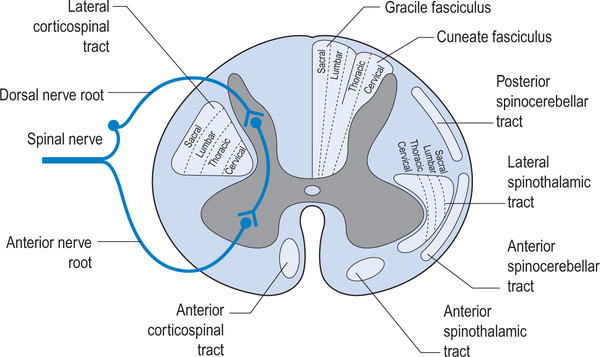
As clinician's, we should be thinking of altered gait as a window to what is going on north of the feet, knees, hip and pelvis. We remember that the joint and muscle mechanoreceptors feedback to the cerebellum and cortex via the spinocerebellar and dorsal column pathways which feed forward to the lower extremities via the anterior spinous cerebellar pathway as well as cortical spinal, rubrospinal had vestibula spinal pathways. The cortex, particularly the motor portion, has the capacity to alter gait just as abnormal mechanoreception has the capacity to alter cortical and cerebellar function. The two are interrelated and inseparable. Changes over time will altered pathways due to neural plasticity and adaptations will occur.

We need to be prudent and examined people fully and be very careful as to the modalities and exercises that we utilize and prescribed as ultimately they will shape that patients neural architecture.


Eggleston JD, Harry JR, Hickman R, Dufek JS. Analysis of gait symmetry during overground walking in children with autism spectrum disorder. Gait Posture 2017;55:162-166. 

Dufek JS, Eggleston JD, Harry JR, Hickman R. A comparative evaluation of gait between children with autism and typically developing matched controls. Med Sci 2017;5:1.  link to free full text: https://www.ncbi.nlm.nih.gov/pmc/articles/PMC5635776/


How about that arm swing?

38846451_2134970409875956_3136689889865629696_n.png

Do you ever notice that sometimes when people have a problem with one of their lower extremities, there are arm swing changes? Usually on the opposite side? This can be anything from a short leg to lack of ankle dorsiflexion, lack of hip extension or even over pronation.

Many times, gait changes, including arm swing, are an "above down" process. This means it begins in the cortex, affects the pattern generators which subsequently will affect extremities distal to that. Sometimes this is a metabolic problem, sometimes vestibular (which can also be mechanical, causing decreased joint and muscle mechanoreceptor input to the cerebellum and vestibular nucleii), sometimes a combination of both. Throw a figure-of-eight ankle wrap on and walk. Your ROM is decreased (mechanical); this reduces input to your cerebellum which reduces input to your vestibular system. Your ankle dorsiflexion and step length will be diminished on that side; this will often cause an increase in arm swing on the contralateral side, which increases the metabolic "cost".

Arm swing may be coached, but we believe this is not always the correct approach as if it is a vestibular problem with altered cerebellar input (Something with the actual semicircular canals or perhaps input from muscle spindle or Golgi tendon organs), coaching arm swing makes the patient "look better" but does not really "fix" the problem; Which may be something as simple as joint pathomechanics, ligamentous restriction or a lack of skilled/endurance/strength in appropriate musculature.  If it is a metabolic issue, sometimes coaching arm swing can improve mechanical efficiency but at the cost of decreasing cortical efficiency, because the brain is such an energy hog.

Arm swing is there for a reason. It tells you something about what is going on or what is not going on. Just because it looks bad does not mean that it is necessarily the problem. Look deeper and keep your eyes, ears and mind open.

We will be talking about the case with this gal. her crossover gait and armswing, alonng with 2 other cases, on our 3rd Wednesdays class on onlince.com: Biomechanics 320  on 8/15/2018

Meyns P, Bruijn SM, Duysens J. The how and why of arm swing during human walking. Gait Posture. 2013 Sep;38(4):555-62. doi: 10.1016/j.gaitpost.2013.02.006. Epub 2013 Mar 13.

Wu Y, Li Y, Liu AM, Xiao F, Wang YZ, Hu F, Chen JL, Dai KR, Gu DY. Effect of active arm swing to local dynamic stability during walking. Hum Mov Sci. 2016 Feb;45:102-9. doi: 10.1016/j.humov.2015.10.005. Epub 2015 Nov 23.

Better gait AFTER rhizotomies?

Nothing surprised me more than reading this paper and finding out that folks that have had rhizotomies, which removes the afferent input from the dorsal horn and sensory information from the reflex loops in the cord, actually had better gait. Of course these children had severe spastic diplegia, which means they have lost descending inhibition from higher center's and most likely had increased flexor tone in the lower extremities. 

image credit: http://realtyconnect.me/spinal-cord-cross-section-tracts/background-information-musculoskeletal-key-within-spinal-cord-cross-section-tracts/

image credit: http://realtyconnect.me/spinal-cord-cross-section-tracts/background-information-musculoskeletal-key-within-spinal-cord-cross-section-tracts/

Remember that the fibers entering the dorsal horn not only go to the dorsal columns but also to the spinocerebellar pathways. When someone has spasticity, the feedback loops are skewed and flexor drive coming from the rostral reticular formation generally is increased are often kept in check by the cerebellar and vestibular feedback loops. Perhaps the interruption of this feedback loop and lack of information from type IA and II afferents of the muscle spindles as well as Ib afferents from the globe tendon organs modulated the tone sufficiently to improve gait. This study did a selective dorsal rhizotomy which means only a portion of it was ablated. 

The somatotopic organization  of the dorsal horn of the spinal cord (i.e.: certain areas of the dorsal horn correspond to certain body parts) is well documented in humans; It would make sense that the dorsal root itself (i.e.: the afferent fibers in the nerve going into the dorsal horn) would be as well, as they are that way in murines (2) and felines (3). 

So, how does this apply to gait? People with strokes, cortical lesions, diseases like cerebral palsy and even possibly increased flexor tone, may benefit from altered input into the dorsal horn. It would have been really cool to see if they increased extensor activity in this individuals, if they would be benefited further. 


Abstract

OBJECTIVE: To identify factors associated with long-term improvement in gait in children after selective dorsal rhizotomy (SDR).

DESIGN: Retrospective cohort study SETTING: University medical center PARTICIPANTS: 36 children (age 4-13y) with spastic diplegia (gross motor classification system level I (n=14), II (n=15) and III (n=7) were included retrospectively from the database of our hospital. Children underwent selective dorsal rhizotomy (SDR) between January 1999 and May 2011. Patients were included if they received clinical gait analysis before and five years post-SDR, age >4 years at time of SDR and if brain MRI-scan was available.

INTERVENTION: Selective dorsal rhizotomy MAIN OUTCOME MEASURES: Overall gait quality was assessed with Edinburgh visual gait score (EVGS), before and five years after SDR. In addition, knee and ankle angles at initial contact and midstance were evaluated. To identify predictors for gait improvement, several factors were evaluated including: functional mobility level (GMFCS), presence of white matter abnormalities on brain-MRI, and selective motor control during gait (synergy analysis).

RESULTS: Overall gait quality improved after SDR, with a large variation between patients. Multiple linear regression analysis revealed that worse score on EVGS and better GMFCS were independently related to gait improvement. Gait improved more in children with GMFCS I & II compared to III. No differences were observed between children with or without white matter abnormalities on brain MRI. Selective motor control during gait was predictive for improvement of knee angle at initial contact and midstance, but not for EVGS.

CONCLUSION: Functional mobility level and baseline gait quality are both important factors to predict gait outcomes after SDR. If candidates are well selected, SDR can be a successful intervention to improve gait both in children with brain MRI abnormalities as well as other causes of spastic diplegia.


1. Oudenhoven LM, van der Krogt MM, Romei M, van Schie PEM, van de Pol LA, van Ouwerkerk WJR, Harlaar Prof J, Buizer AI. Factors associated with long-term improvement of gait after selective dorsal rhizotomy. Arch Phys Med Rehabil. 2018 Jul 4. pii: S0003-9993(18)30442-8. doi: 10.1016/j.apmr.2018.06.016. [Epub ahead of print]

2. Wessels WJ1, Marani E. A rostrocaudal somatotopic organization in the brachial dorsal root ganglia of neonatal rats. Clin Neurol Neurosurg. 1993;95 Suppl:S3-11.

3. Koerber HRBrown PB. Somatotopic organization of hindlimb cutaneous nerve projections to cat dorsal horn. J Neurophysiol. 1982 Aug;48(2):481-9.

Headbonking and gait

A great article (see reference below) just came out looking at the gait changes that come along with a concussion. Basically it says that folks that have concussions have more coronal plane (i.e. side to side) sway and they walk slower. This reminded us of some of the "decomposition of gait" pieces that we have done and one post on proprioceptive clues in children gait that we did about 5 years ago. Having a concussion causes decomposition of gait, and we move toward a more primitive pattern, just like we see in kids. Here was the post:

We can learn a lot about gait from watching our children walk. An immature nervous system is very similar to one which is compensating meaning cheating around a more proper and desirable movement pattern; we often resort to a more primitive state when challenges beyond our ability are presented. This is very common when we lose some aspect of proprioception, particularly from some peripheral joint or muscle, which in turn, leads to a loss of cerebellar input (and thus cerebellar function). Remember, the cerebellum is a temporal pattern generating center so a loss of cerebellar sensory input leads to poor pattern generation output. Watch this clip several times and then try and note each of the following:

  • wide based gait; this is because proprioception is still developing (joint and muscle mechanoreceptors and of course, the spino cerebellar pathways and motor cortex)
  • increased progression angle of the feet: this again is to try and retain stability. External rotation allows them to access a greater portion of the glute max and the frontal plane (engaging an additional plane is always more stable).
  • shortened step length; this keeps the center of gravity close to the body and makes corrections for errors that much easier (remember our myelopathy case from last week ? LINK.  This immature DEVELOPING system is very much like a mature system that is REGRESSING.  This is a paramount learning point !)
  • decreased speed of movement; this allows more time to process proprioceptive clues, creating accuracy of motion

Remember that Crosby, Still, Nash and young song “Teach Your Children”? It is more like, “teach your parents”…

Proprioceptive clues are an important aspect of gait analysis, in both the young and old, especially since we tend to revert back to an earlier phase of development when we have an injury or dysfunction.


Manaseer TSGross DPDennett LSchneider KWhittaker JL1. Gait Deviations Associated With Concussion: A Systematic Review.  Clin J Sport Med. 2017 Nov 21. doi: 10.1097/JSM.0000000000000537. [Epub ahead of print]

Party over the Weekend?


So, the more you drink, the more you impair the CNS. The more you impair the CNS, the more dependent you become on peripheral mechanisms. A good reason to keep your vestibular system (alcohol changes the specific gravity of the endolymph), your visual system (long term use affects the option nerve directly and can cause involuntary saccades) and proprioceptive systems including the cutaneous mechanoreceptors (because you are relying on them more) intact. Watch what and how much you drink...

"Standing postural stability relies on input from visual, vestibular, proprioceptive and mechanoreceptive sensors. When the information from any of these sensors is unavailable or disrupted, the central nervous system maintains postural stability by relying more on the contribution from the reliable sensors, termed sensory re-weighting. Alcohol intoxication is known to affect the integrity of the vestibular and visual systems. The aim was to assess how mechanoreceptive sensory information contributed to postural stability at 0.00% (i.e. sober), 0.06% and 0.10% blood alcohol concentration (BAC) in 25 healthy subjects (mean age 25.1 years). The subjects were assessed with eyes closed and eyes open under quiet standing and while standing was perturbed by repeated, random-length, vibratory stimulation of the calf muscles. Plantar cutaneous mechanoreceptive sensation was assessed for both receptor types: slowly adapting (tactile sensitivity) and rapidly adapting (vibration perception). The correlation between recorded torque variance and the sensation from both mechanoreceptor types was calculated. The recorded stability during alcohol intoxication was significantly influenced by both the tactile sensation and vibration perception of the subjects. Moreover, the study revealed a fluctuating association between the subjects' vibration perception and torque variance during balance perturbations, which was significantly influenced by the level of alcohol intoxication, vision and adaptation. Hence, one's ability to handle balance perturbations under the influence of alcohol is strongly dependent on accurate mechanoreceptive sensation and efficient sensory re-weighting. 

Modig F, Patel M, Magnusson M, Fransson PA.Study II: mechanoreceptive sensation is of increased importance for human postural control under alcohol intoxication. Gait Posture. 2012 Mar;35(3):419-27. doi: 10.1016/j.gaitpost.2011.11.001. Epub 2011 Dec 27.
weighting.