One way compensations develop
We have all had injuries; some acute some chronic. Often times injuries result in damage to the joint or articulation;  when the ligament surrounding a joint becomes injured we call this a “sprain”. 
Joints are blessed …

One way compensations develop

We have all had injuries; some acute some chronic. Often times injuries result in damage to the joint or articulation;  when the ligament surrounding a joint becomes injured we call this a “sprain”. 

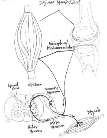
Joints are blessed with four types of mechanoreceptors.  We have covered this in many other posts (see here and here).  These mechanoreceptors apprise the central nervous system of the position (proprioception or kinesthesis) of that body part or joint via the dorsal column system or spinocerebellar tracts. Damage to these receptors can result in a mismatch or inaccuracy of information to the central nervous system (CNS). This can often result in further injury or a new compensation pattern. 

Joints have another protective mechanism called arthrogenic inhibition (see diagram above). This protective reflex turns off the muscles which cross the joint. This was described in a few great paper by Iles and Stokes in the late 80’s an early 90’s (vide infra). Not only are the muscles inhibited, but it can also lead to muscle wasting; there does not need to be pain and a small joint effusion can cause the reflex to occur. 

If the muscles are inhibited and cannot provide appropriate afferent (sensory) and efferent (motor) information to the CNS, your brain makes other arrangements to have the movement occur, often recruiting muscles that may not be the best choice for the job. We call this a “compensation” or “compensation pattern”. An example would be that if the glute max is inhibited (a 2 joint muscle, with a larger attachment to the IT band and a smaller to the gluteal tuberosity; it is a hip extender, external rotator and adductor of the thigh), you may use your lumbar erectors (multi joint muscles; extensors and lateral rotators of the lumbar spine) or hamstrings (2 joint muscles; hip extenders, knee flexors, internal and external rotators of the thigh)  to extend the hip on that side, resulting in aberrant mechanics often observable in gait, which may manifest itself as a shortened step length, increased vertical displacement of the pelvis, lateral shift of the pelvis or increase in step height, just to name a few. Keep this up for a while and the new “pattern” becomes ingrained in the CNS and that becomes your new default for that motion.

Now to fix the problem, you not only need to reactivate the muscle, but you need to retrain the activity. Alas, the importance of doing a thorough exam and thorough rehab to fix the problem.

Often times, the fix is much more involved than figuring out what the problem is (or was). Take your time and do a good job. Your clients and patients will appreciate it!

Ivo and Shawn, the gait guys

Young A, Stokes M, Iles JF : Effects of joint pathology on muscle. Clin Orthop Relat Res. 1987 Jun;(219):21-7

Iles JF, Stokes M, Young A.: Reflex actions of knee joint afferents during contraction of the human quadriceps. Clin Physiol. 1990 Sep;10(5):489-500.

image from: http://chiroeco.com/chiro-blog/results-to-referrals/2013/04/03/neurology-based-simplified-musculoskeletal-assessment/

Functional Ankle Instability and the Peroneals  

Lots of links available here with today’s blog post. please make sure to take your time and check out each one (underlined below)  
As you remember, the peroneii (3 heads) are on the outside of…

Functional Ankle Instability and the Peroneals 


Lots of links available here with today’s blog post. please make sure to take your time and check out each one (underlined below) 

As you remember, the peroneii (3 heads) are on the outside of the lower leg (in a nice, easy to remember order of longus, brevis and tertius, from top to bottom) and help to stabilize the lateral ankle. The peroneus brevis and tertius dorsiflex and evert the foot while the peroneus longus plantarflexes and everts the foot. We discuss the peroneii more in depth here in this post. It then is probably no surprise to you that people with ankle issues, probably have some degree of peroneal dysfunction. Over the years the literature has supported notable peroneal dysfunction following even a single inversion sprain event. 

Functional ankle instability (FAI) is defined as “ the subjective feeling of ankle instability or recurrent, symptomatic ankle sprains (or both) due to proprioceptive and neuromuscular deficits." 

Arthrogenic muscle inhibition (AMI) is a neurological phenomenon where the muscles crossing a joint become "inhibited”, sometimes due to effusion (swelling) of the joint (as seen here) and that may or may not be the case with the ankle (see here), or it could be due to nociceptive input altering spindle output or possibly higher centers causing the decreased muscle activity. 

This paper (see abstract below) merely exemplifies both the peroneals and FAI as well as AMI.

Take home message?

Keep the peroneals strong with lots of balance work!

The Gait Guys: bringing you the meat, without the filler!                                                                         

Am J Sports Med. 2009 May;37(5):982-8. doi: 10.1177/0363546508330147. Epub 2009 Mar 6.

Peroneal activation deficits in persons with functional ankle instability.

Source

School of Kinesiology, University of Michigan, 401 Washtenaw Avenue, Ann Arbor, MI 48109, USA. riannp@umich.edu

Abstract

BACKGROUND:

Functional ankle instability (FAI) may be prevalent in as many as 40% of patients after acute lateral ankle sprain. Altered afference resulting from damaged mechanoreceptors after an ankle sprain may lead to reflex inhibition of surrounding joint musculature. This activation deficit, referred to as arthrogenic muscle inhibition (AMI), may be the underlying cause of FAI. Incomplete activation could prevent adequate control of the ankle joint, leading to repeated episodes of instability.

HYPOTHESIS:

Arthrogenic muscle inhibition is present in the peroneal musculature of functionally unstable ankles and is related to dynamic peroneal muscle activity.

STUDY DESIGN:

Cross-sectional study; Level of evidence, 3.

METHODS:

Twenty-one (18 female, 3 male) patients with unilateral FAI and 21 (18 female, 3 male) uninjured, matched controls participated in this study. Peroneal maximum H-reflexes and M-waves were recorded bilaterally to establish the presence or absence of AMI, while electromyography (EMG) recorded as patients underwent a sudden ankle inversion perturbation during walking was used to quantify dynamic activation. The H:M ratio and average EMG amplitudes were calculated and used in data analyses. Two-way analyses of variance were used to compare limbs and groups. A regression analysis was conducted to examine the association between the H:M ratio and the EMG amplitudes.

RESULTS:

The FAI patients had larger peroneal H:M ratios in their nonpathological ankle (0.399 +/- 0.185) than in their pathological ankle (0.323 +/- 0.161) (P = .036), while no differences were noted between the ankles of the controls (0.442 +/- 0.176 and 0.425 +/- 0.180). The FAI patients also exhibited lower EMG after inversion perturbation in their pathological ankle (1.7 +/- 1.3) than in their uninjured ankle (EMG, 3.3 +/- 3.1) (P < .001), while no differences between legs were noted for controls (P > .05). No significant relationship was found between the peroneal H:M ratio and peroneal EMG (P > .05).

CONCLUSION:

Arthrogenic muscle inhibition is present in the peroneal musculature of persons with FAI but is not related to dynamic muscle activation as measured by peroneal EMG amplitude. Reversing AMI may not assist in protecting the ankle from further episodes of instability; however dynamic muscle activation (as measured by peroneal EMG amplitude) should be restored to maximize ankle stabilization. Dynamic peroneal activity is impaired in functionally unstable ankles, which may contribute to recurrent joint instability and may leave the ankle vulnerable to injurious loads.

all material (except for the study); copyright 2013 The Gait Guys/ The Homunculus Group. All rights reserved. Please ask before you lift our stuff. If you are nice and give us credit, we will probably let you use it!