Which foot exercises activate the intrinsics?

So, your goal is to strengthen the intrinsics. What exercise is best? Probably the most specific one, right? Well....maybe. These 4 exercises seem to all hit them.

This study looked at the muscle activation of the abductor hallucis, flexor digitorum brevis, abductor digiti minimi, quadratus plantae, flexor digiti minimi, adductor hallucis oblique, flexor hallucis brevis, and interossei and lumbricals with the short foot, toe spreading, big toe extension and lesser toes extension exercises with T2 weighted MRI post exercises (perhaps not the best way to look at it) and shows they all work to varying degrees.

"All muscles showed increased activation after all exercises. The mean percentage increase in activation ranged from 16.7% to 34.9% for the short-foot exercise, 17.3% to 35.2% for toes spread out, 13.1% to 18.1% for first-toe extension, and 8.9% to 22.5% for second- to fifth-toes extension."

Gooding TM, Feger MA, Hart JM, Hertel J. Intrinsic Foot Muscle Activation During Specific Exercises: A T2 Time Magnetic Resonance Imaging Study. Journal of Athletic Training. 2016;51(8):644-650. doi:10.4085/1062-6050-51.10.07.

link to full text: https://www.ncbi.nlm.nih.gov/pmc/articles/PMC5094843/

tumblr_nhbuqbFrTD1qhko2so1_250.png
tumblr_nhbuqbFrTD1qhko2so2_500.png
tumblr_nhbuqbFrTD1qhko2so3_r1_1280.jpg

When the big guy heads medially….Game Changer

Lately we have been seeing a lot of bunions (hallux valgus). While doing some research on intermetatarsal angles (that’s for another post) we came across the nifty diagram you see above. 

Regardless of the cause, as the 1st metatarsal moves medially, there are biomechanical consequences. Lets look at each in turn. 

  • the EHB (extensor hallucis brevis) axis shifts medially. this muscle, normally an extensor of the proximal phalanyx, now becomes more of an abductor of the hallux. It’s secondary action of assisting the descent of the head of the 1st metatarsal no longer happens and it actually moves the base of the proximal phalanyx posteriorly, altering the axis of centration of the joint, contributing to a lack of dorsiflexion of the joint and a hallux limitus
  • Abductor hallucis becomes more of a flexor, as it moves to the plantar surface of the foot. Remember, a large percentage of people already have this muscle inserting more on the plantar surface of the foot (along with the medial aspect of the flexor hallucis brevis), so in these folks, it moves even more laterally, distorting the proximal phalanx along its long axis (ie medially) see this post here for more info
  • Flexor hallucis brevis moves more laterally. Remember this muscle houses the sesamoid bones before inserting onto the base of the proximal phalannx; the medial blending with the abductor hallucis and the lateral with the adductor hallucis. Because the sesamoid bones have moved laterally, they no longer afford this muscle the mechanical advantage they did previously and the axis of motion of the 1st metatarsal phalangeal joint moves dorsally and posterior, contributing to limited dorsiflexion of that joint and a resultant hallux limitis. The lateral movement of the sesamoids also tips the long axis of the 1st metatarsal and proximal phalanyx into eversion. In addition, the metatarsal head is exposed and is subject to the ground reactive forces normally tranmittted through the sesamoids; often leading to metatarsalgia. 
  • Adductor hallucis: this muscle now has a greater mechanical advantage  and because the head of the 1st ray is not anchored, acts to abduct the hallux to a greater degree. The now everted position of the hallux contributes to this as well

As you can see, there is more to the whole than the sum of the parts. Bunions have many biomechanical consequences, and these are only a small part of the big picture. Take you time, learn your anatomy and examine everything that has a foot!

See you in the shoe isle…

Ivo and Shawn

pictures from: http://www.orthobullets.com/foot-and-ankle/7008/hallux-valgus and http://www.stepbystepfootcare.com/faqs/nakedfeet/

tumblr_my9vb37Ozy1qhko2so1_1280.jpg
tumblr_my9vb37Ozy1qhko2so2_1280.jpg
tumblr_my9vb37Ozy1qhko2so3_540.png

And what have we here?

The above is a pedograph, a simple, effective pressure map of the foot as someone is walking across an inked grid. For more info on pedographs, click here.

Did you note the increased ink present under the great toe bilaterally? What could be causing this? If you look carefully, you will note that it is at the base of the proximal phalynx of the great toe. This could be none other than the tendon of the flexor hallucis brevis!

This bad boy arises from the medial part of the under surface of the cuoid and the adjacent 3rd cunieform, with a small slip from the tendon of the tibialis posterior. As it travels forward it splits into two parts, which are inserted into the medial and lateral sides of the base of the proximal phalanyx of the great toe. There is a sesamoid bone present in each tendon, which offers the FHB a mechanical advantage when flexing the toe.  The medial portion is blends with the abductor hallucis and the lateral portion blends with the adductor hallucis.

Had the increased printing on the pedograph been more distal, it most likely would have been due to increased action of the flexor hallucis longus.   Had it been more proximal (under the head of the 1st metatarsal) it would have been due to the peroneus longus.

Cool, eh?

Reading pedographs and making you a sharper clinician/coach/trainer/sales person is just one of the many skills we try to teach here on the blog. Keep up the great work!

The Gait Guys


Why alignment of the big toe is so critical to gait, posture, stabilization motor patterns and running.

There are two ways of thinking about the arch of the foot when it comes to competent height.  One perspective is to passively jack up the arch with a device such as an orthotic, a choice that we propose should always be your last option, or better yet to access the extrinsic and intrinsic muscles of the foot (as shown in this video) to compress the legs of the foot tripod and lift the arch dynamically.  Here today we DO NOT discuss the absolute critical second strategy of lifting the arch via the extensors as you have seen in our “tripod exercise video” (link here) but we assure you that regaining extensor skill is an absolute critical skill for normal arch integrity and function.  We like to say that there are two scenarios going on to regain a normal competent arch (and that does not necessarily mean a high arch, a low arch can also be competent….. it is about function and less about form): one scenario is to hydraulically lift the arch from below and the other scenario is to utilize a crane-like effect to lift the arch from above. When you combine the two, you restore the arch function.  In those with a flat flexible incompetent foot you can often regain normal alignment and function.  But remember, you have to get to the client before the deforming forces are significant enough and have been present long enough that the normal anatomical alignments are no longer possible. For example, a hallux valgus with a large bunion (this person will never get to the abductor hallucis sufficiently) or a progressively collapsed arch that is progressively becoming rigid or semi-rigid.

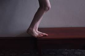
Think about these concepts today as you watch your clients walk, run or exercise.  And then consider this study below on the critical importance of the abductor hallucis muscle after watching our old video of Dr. Allen’s competent foot.  

CONCLUSIONS AND CLINICAL RELEVANCE:

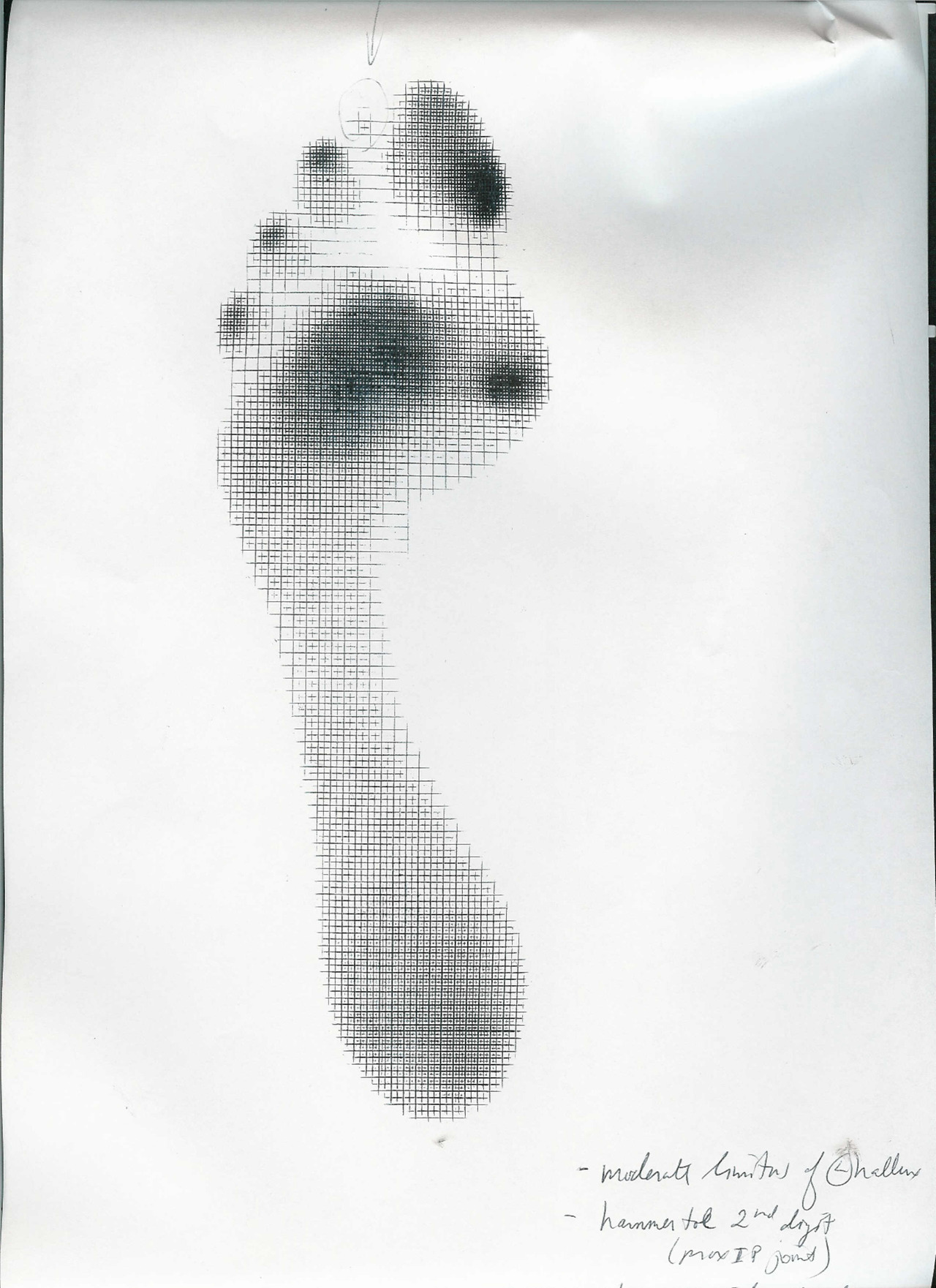
The abductor hallucis muscle acts as a dynamic elevator of the arch. This muscle is often overlooked, poorly understood, and most certainly rarely addressed. Understanding this muscle and its mechanics may change the way we understand and treat pes planus, posterior tibial tendon dysfunction, hallux valgus, and many other issues that lead to a challenge of the arch, effective and efficient gait. Furthermore, its dysfunction and lead to many aberrant movement and stabilization strategies more proximally into the kinetic chains.

*From the article referenced below,  “Most studies of degenerative flatfoot have focused on the posterior tibial muscle, an extrinsic muscle of the foot. However, there is evidence that the intrinsic muscles, in particular the abductor hallucis (ABH), are active during late stance and toe-off phases of gait.“

We hope that this article, and the video above, will bring your focus back to the foot and to gait for when the foot and gait are aberrant most proximal dynamic stabilization patterns of the body are merely strategic compensations.

Study RESULTS:

All eight specimens showed an origin from the posteromedial calcaneus and an insertion at the tibial sesamoid. All specimens also demonstrated a fascial sling in the hindfoot, lifting the abductor hallucis muscle to give it an inverted ‘V’ shaped configuration. Simulated contraction of the abductor hallucis muscle caused flexion and supination of the first metatarsal, inversion of the calcaneus, and external rotation of the tibia, consistent with elevation of the arch.

http://www.ncbi.nlm.nih.gov/pubmed/17559771

Foot Ankle Int. 2007 May;28(5):617-20.

Influence of the abductor hallucis muscle on the medial arch of the foot: a kinematic and anatomical cadaver study.

Wong YS. Island Sports Medicine & Surgery, Island Orthopaedic Group, #02-16 Gleneagles Medical Centre, 6 Napier Road, Singapore, 258499, Singapore. 

Using a Pedograph to get Dynamic Answers to Foot Dysfunction:
Pedograph topic: One quick topic here. Note the hot spot (ink concentration) at the big toe and note that the ink is proximal on the toe pad. We would like to see the pressure point at th…

Using a Pedograph to get Dynamic Answers to Foot Dysfunction:

Pedograph topic: One quick topic here. Note the hot spot (ink concentration) at the big toe and note that the ink is proximal on the toe pad. We would like to see the pressure point at the center of the pad. This spot means this person walked across the Harris Ink mat with increased FHB (flexor hallucis brevis) use and not enough FHL (flexor hallucis longus);  too much short flexor, not enough long flexor. There is loss of synergy between the two. This will likely mean there is something going on in the extensors as well, something abnormal.

Need a review? Look at Monday’s video again on the EHB (extensor hallucis brevis) where we discuss all of the toes muscles.

Clinically this patient had a hallux limitus/rigidus (could not dorsiflex great toe) which complicated the mechanics at the joint and forward into the great toe, sadly also at the foot’s medial tripod as well. You cannot get an accurate read from a static (standing only) pressure mapping. Don’t rely on them for dynamic info !

Too much FHB with not enough FHL means EHB (as well as long extensors of the lesser toes) is going to be impaired. Impair the EHB and you ask the EHL  to work differently as well.  Here’s a hint, look at all the printing under the lesser digits distally, there is too much flexor activity here as indicated by intense inking from toe clenching / hammering.  They are likely doing this to add more stability since the great toe cannot from what we discussed above. There are problems that come from these issues as well but we want to stay focused on the big toe today.

Now, go back and review Mondays blog video post (here is the link).

Treatment:

In a case where there is some loss of the 1st MPJ range of motion (metatarsaphalangeal joint) (depending on the source, 45 degrees is typically needed) there will be impairment of the long and short toe flexor/extensor pairing and synergy.  In this case above there is highly suspected increased short flexor (FHB) activity (hence the ink at the proximal big toe) and this means that the long flexor is usually submissive.  And, when the long flexor (FHL) is submissive the long extensor is dominant. When the long extensor is dominant the short extensor is submissive. Can you now see the beautiful symphony and harmony we need here. This is why we loosely say that the FHB and the EHL are paired and the FHL and EHB are paired.  It is not exactly the case but hopefully you catch our drift. 

So, in this case, with a hallux limitus/rigidus when the 45 degrees of dorsiflexion is lost these pairing can be challenges and the synergy is lost.  The symphony of these muscles is “off tune”.  This can further provoke the 1st MPJ and it can also be the slow brewing initiation of the problem. It can be a vicious cycle when it gets going. And, when the 1st MPJ is limited the dorsiflexion that is supposed to occur at the joint can be shunted proximally into the midfoot or ankle and cause pain/pathology there.  It can also impair the normal pronation-supination cycles. The big toe when it goes sour makes the whole orchestra angry and play off tune.

Doing your best to normalize and maximize muscle harmony and function many times will dampen the pathology and pain and get the person going again.  Of course the problem is still lurking under the surface.  Test the muscles, try to isolate them but remember that your muscle tests need to be as specific as you can. Nothing is isolated in the body, but do your best.

Of course there are many other scenarios but this is the one we chose to teach today from this pedographing of the big toe. We will explore other options and challenges another time.

Shawn and Ivo.    Gait geeks promoting gait literacy and competency everywhere we can get an open ear.

 Splay

Watch this video a few times through. Did you catch the subtle abduction moment of the Hallux (big toe) on impact? Did you see the collapse of the transverse metatarsal arch? No?  Watch it until you do.

What gives? We thought toes were supposed to be stable when they hit the ground (and in fact they are).  Read on…

Think of the adductor hallucis. It has 2 heads. The oblique head arises from the proximal shafts of metatarsals 2-4 and inserts on the MEDIAL aspect of the proximal phalynx of the hallux (along with medial fibers of the flexor hallucis brevis); the transverse head arises from the metatarsophalangeal ligaments of  digits 3-5, and the transverse metatarsal ligament and inserts blending with the oblique head on the proximal phalynx of the hallux.

The action of the adductor hallucis mirrors that of the abductor hallucis (which inserts on the LATERAL side of the proximal phalynx. Together, they act to keep the hallux straight and provide a compressive force which stabilizes the big toe WHEN IT IS ON THE GROUND.

The problem here, is that the base of the Hallux is NOT anchored to the ground. This person has a faulty tripod (most likely an uncompensated forefoot varus) and cannot anchor the big toe, there fore the adductor cannot do it’s job. Is is weak (from lack of use) and we see the result: an abducting big toe AND collapse of the transverse metatarsal arch (which the transverse head of the adductor, under normal conditions maintains).

Looks like this guy needs some exercises to descend the head of the 1st metatarsal and make an adequate tripod. Flexing the distal phalynx of the hallux while extending the metatarsophalangeal joint would be a good start. (see Dr Allen demonstrate this here: http://www.youtube.com/user/TheGaitGuys?feature=grec_index#p/u/11/TyRE9dReVTE )

The Gait Guys…promoting foot literacy here and everywhere.

The Gait Guys: Some strategies in Controlling the Foot Arches and Big Toe

As promised. We fixed the volume.  Less hiss next time. Enjoy

Dr. Shawn Allen of The Gait Guys speaks about proper stabilization of the medial foot and arch. Muscle specifically discussed are a team: FHB (flexor hallucis brevis), AbDuctor hallucis, and tibialis posterior. He discusses the functional anatomy, normal and pathologic movement patterns of the arch and first ray complex and big toe (hallux). His foot’s ability to show the optimal patterns for the arch and hallux are excellent examples. Follow up videos and DVDs will show more details you need to know, and some of the exercises he and Dr. Ivo Waerlop use to restore a foot that has lost these abilities. The DVDs are in the works. Take their lectures and CME on www.onlineCE.com. Visit them at www.thegaitguys.com and on their facebook PAGE & Twitter of the same name for daily feeds of unique things.