The importance of hip extension


Watch this clip a few times. Pause it when it pauses. Have a look, then continue. Come back here when you are done.


Sometimes it seems like we keep saying the same thing over and over. Remember the 3 rockers? Heel rocker, ankle rocker and forefoot rocker. Look at the clip again. All three are there. We stopped the clip in a few places to show you HOW MUCH ankle rocker there was; probably a little TOO MUCH. As for hip extension, on the exam table, this gal had at least 15 degrees of hip extension. So what gives?


All that is lack of hip extension is not loss of ankle rocker, as you can see here. If we don’t have hip extension (she never even gets to zero) , then something else needs to do the brunt of the work. Here we see her quads and calves getting quite the workout here. But look closer and watch the lumbar lordosis (curve in the lumbar spine). See how it stiffens on impact and then extends as she goes through toe off? The motion HAS to occur SOMEWHERE. Guess we know why she has low back pain, especially on long runs.


The take home message?


We need 3 competent rockers for normal gait

We need adequate hip extension for normal gait

When one of the above mechanisms is not in place, compensation will occur somewhere else in the chain


The fix? Train her to use her hip extensors (glue max and medius) through gluteal awareness exercises. Things like having her palpate her glutes while walking and running.  Texas walk exercises. Retrain her foot strike to be more positioned under her body. Get her to extend and bring her arms back more. Stuff we talk about here all the time.


Ivo and Shawn. The Gait Guys. Extending your cerebral hips each and every post.


All material copyright 2013 The Gait Guys/ The Homunculus Group.

Some honest movements across the big toe. Things you need to know as a runner or walker.

Lets see how good you are at this gait game at this point. 

In the video above you should see two things: you should see me manually dorsiflexing / extending the big toe / hallux.  I am creating extension through the 1st metatarsal joint (1st MTP). Essentially I am passively engaging the Windlass Mechanism of Hicks.  This mechanism is essentially a biomechanical event that wraps the end of the plantar fascia over the metatarsal head and 1st MTP joint.  So, when a person raises the heel in gait a moment of dorsiflexion/extension occurs across the joint.  In the video you see me lifting the toe but in the closed chain event the toe stays on the ground and foot moves up and over the toe but the resultant motion at the 1st MTP is the same.  It is still dorsiflexion / extension of the big toe about the 1st MTP joint. 
This wrapping or winding of the plantarfascia around the joint causes the distance from the heel to the ball of the foot to shorten and thus creates an elevation of the arch of the foot (smarter than using an orthotic to push it up  huh !) but this mechanism also raises the talus and supinates the foot.  This action makes the foot more rigid and stable. After all, when you are raising the heel and progressing over the ball of the foot don’t you want a rigid lever to press off of ?!  Ask any sprinter and they will concur. However, this mechanism occurs in all folks who  have a relatively competent foot and 1st MTP joint. 
What you NEED to see in the video is the additional motion up the limb. Watch the video again. When the toe is extended (dorsiflexed) the arch rises but the limb also externally rotates. You can see this by the subtle drift of the blue dots on the limb.  We want and need this external rotation to occur at the hip and through the limb because remember, the limb was internally rotated as we passed our body mass over the foot. This is a normal gait phenomenon.
Here is what we want to you to ponder.  Imagine a person with:

  •  a weak extensor hallucis mechanism (both longus and brevis: EHL, EHB)
  •  a hallux rigidus where the toe does not fully extend to that magical 45+ degree range or
  • if the arch of the foot is so far collapsed and pronated

IF any of these things go wrong, then a sufficiently rigid foot is not formed for push off propulsion, an incompetent arch results and insufficient talar motion and external limb rotation will occur. This means that several subsequent biomechanical events will/ may be compromised including but not limited to:

  • contralateral arm swing
  • sufficient engagement of the gluteals for stablization and propusion since optimal external hip rotation will not be met
  • inadequate pelvis posturing for spine neutrality 
  • etc. this is potentially a very very long list

Our take home point here is simple. You must have:

  •  sufficient and relatively symmetrical bilateral big toe extenion (hallux dorsiflexion) to optimize the Windlass Effect
  • you must have sufficient strength of the toe extensors to gain and stabilize this joint range at the 1st MTP joint
  • you must have both of the prior 2 in order to properly posture the foot and arch for rigid terminal loading response
  • and you must have all 3 to sufficiently externally rotate the limb at the hip to engage the propulsive components of the gait cycle.

There is so much more we could have added to this brief blog post, including hallux impingement, hallux valgus and bunion formation, supination insufficiency syndromes, tibialis posterior insufficiency syndrome, metatarsalgia etc.  The list is endless. 

Hallux dorsiflexion, both passive and active range of motion must be checked on every athlete and client you see. Otherwise gait is likely to be impaired locally and globally from subtle insufficiencies.

Shawn and Ivo
The Gait Guys

Podcast #25: Bionics, Arm Swing & Footwear

Great podcast today, #25. Wide range of topics today: the first truly bionic body part, technical shoe issues, GTO’s and more. 

podcast link: 

http://thegaitguys.libsyn.com/podcast-25-bionics-arm-swing-footwear

iTunes link: https://itunes.apple.com/us/podcast/the-gait-guys-podcast/id559864138

Gait Guys online /download store:

http://store.payloadz.com/results/results.aspx?m=80204

Today’s show notes:


1. The First Truly Bionic Hand

http://www.independent.co.uk/life-style/gadgets-and-tech/news/a-sensational-breakthrough-the-first-bionic-hand-that-can-feel-8498622.html

“The first bionic hand that allows an amputee to feel what they are touching will be transplanted later this year in a pioneering operation that could introduce a new generation of artificial limbs with sensory perception.

2. Effects of toning shoes on lower extremity gait biomechanics

http://www.clinbiomech.com/article/S0268-0033%2813%2900010-7/abstract

Clinical Biomechanics, Jan 2013

3. Beware of trendy barefoot running shoes - you could end up with broken bones in your foot

http://www.dailymail.co.uk/health/article-2289725/Beware-trendy-barefoot-running-shoes–end-broken-bones-foot.html?ito=feeds-newsxml

  • Advocates of barefoot running claim it can reduce injuries and back pain
  • ‘Minimalist’ shoes such as these now account for 15% of sales
  • But experts say many people suffer injuries by overdoing it early on
  • Runners should make transition from regular trainers more slowly, they say

4. Foot strike and injury rates in endurance runners: a retrospective study.
Daoud AI, Geissler GJ, Wang F, Saretsky J, Daoud YA, Lieberman DE.
Med Sci Sports Exerc. 2012 Jul;44(7):1325-34. doi: 10.1249/MSS.0b013e3182465115.

Department of Human Evolutionary Biology, Harvard University, Cambridge, MA 02138, USA.

5. Effects of foot strike on low back posture, shock attenuation, and comfort in running.

http://www.ncbi.nlm.nih.gov/m/pubmed/23073217/
Med Sci Sports Exerc. 2013 Mar;45(3):490-6

CONCLUSION: Change in foot strike from RFS to FFS decreased overall ROM in the lumbar spine but did not make a difference in flexion or extension in which the lumbar spine is positioned. Shock attenuation was greater in RFS. RFS was perceived a more comfortable running pattern.

*it seems to becoming a question as to what you are doing with the body parts at impact……..where it be you are RFS or FFS.  Do you have the ability to protect the parts in varying mechanical stressful positions.

6. Hey guys, Dr. Ryan:

I just listened to Pod 23 and Ivo you mentioned sagittal curves not developing until after birth..  There is evidence they begin to develop in-utero.  Here is an article excerpt and link to it.
 
"In many anatomy texts, it is often claimed and/or assumed that the cervical lordosis is a secondary curve and is not present during intra-uterine life. However, as early as 1977, Bagnall et al3 demonstrated that the cervical lordotic curve is formed in intrauterine life (9.5 weeks). In 195 fetuses, Bagnall et al3 found that by 9.5 weeks, 83% of fetuses have a cervical lordosis, 11% have a military configuration, and only 6% of fetuses are in the typically described kyphotic position of the cervical spine. This means that by 9.5 weeks, 94% of the fetuses are starting to use their posterior cervical muscles to pull the cervical curve away from the fetal “C”-shape. Fetuses have a cervical lordosis before birth, however, the lordosis increases during post-natal life at ages 3 months-9 months as the infant raises his/her head and begins to sit up.4”

REFERENCES

  1. Harrison DD, et al. Spine 1996; 21: 667-675.
  2. Harrison DD, et al. Spine 2004; 29:2485-2492.
  3. Bagnall KM, et al. J Anat 1977;124:791-802.
  4. Kure S. J Tokyo Med Collage 1972;30;453-470.
  5. Kasai T, et al. Growth. Spine 1996;21:2067-2073.
  6. Harrison DE, Harrson DD, Haas JW. Evanston, WY: Harrison CBP Seminars, Inc., 2002, ISBN 0-9721314-0-X.
  7. Shatz A, et al. Acta Anat 1994;149:141-145.
  8. McAviney J, et al. J Manipulative Physiol Ther 2005;28:187-193.
  9. Bastecki A, et al. ADHD: A CBP Case Study. J Manipulative Physiol Ther 2004; 27(8):e14.


7. “Dynamic Arm Swing in Human Walking, (http://www.ncbi.nlm.nih.gov/pubmed/19640879) where it was determined that normal arm swinging required minimal shoulder torque, while volitionally holding the arms still required 12% more metabolic energy. Among measures of gait mechanics, vertical ground reaction moment was most affected by arm swinging and increased by 63% without it.
* brings up issues of shoulder pathology……rot cuff, frozen shoulder, carrying a purse, water bottle etc


8. Winter foot wear:
We like Steger Mukluks…….youtube video   "gait guys mukluks”

9. Versions: one of the more difficult concepts to grasp…………..here is a Q from a FB reader

  • Does retroversion mean this child will automatically grow up with abnormal mechanics - leading to possible knee foot hip back issue etc? Is there a fix to prevent such without an ortho’s bone saw?
     
    10. The role of GTO’s in plyometric exercises.

“Those guys are perfect examples of pure genius.” - Mikhail Baryshnikov

***** WAIT ! Read the blog post FIRST, then watch the clip. Trust us.

We are going to start today’s blog post with a disclaimer.  “Do not attempt what these fellas do in the last moments of this video, particularly the scene on the stairs.”

Almost everyone on the face of this planet can walk, and most of those can also run as well.  It is basically all about putting one foot in front of the other and trying to maintain some sense of balance and stability over the stance limb without falling over. For some however, this is their greatest challenge of the day, walking.  Whether it be from an arthritic hip or knee or a neuro-degenerative disease, some folks see walking as their greatest physical challenge on a daily basis. 

For the able bodied folks, dance is another matter when compared to walking or running. Dance is about as far in the extreme opposite direction as one can get from simple walking gait or running. Here at the Gait Guys we know this intimately. In our mission to better understand human locomotion we continue to personally delve into tasks of complex motion, for it is only through studying the difficult that the beauty of the simple shines through.  After committing 3 years to investigating and learning smooth and latin dance with some truly amazing teachers we can say with some strong personal conviction, dance is different.  Footwork can be very complex in dance, as it can at times in many sports, but one thing is for certain they are not the same.  In dance the foot steps are consciously calculated to the beat of the music, this does not occur in any other sport and thus the steps and lower limb movements in most sport are less calculated and important than when it comes to fixed techniques, procedures and protocols as in dance.  Rumba steps are different from cha cha, waltz, foxtrot, swing, salsa, mambo, hustle, tango etc. Each dance has unique steps and must be able to be performed at varying tempos, at the very least. Oh, least we forget to mention that you usually have a partner you must choreograph the movements with, taking turns moving forward, backward or spinning. In contrast, when Michael Jordan is spinning off of a pick-and-roll driving to the hoop he is not exactly consciously calculating footwork at a ¾ time for exampleor making sure that there was a specific foot and leg action that was premised on the movement. The goal and demand is different in dance. 

There are no particular learning issues on this blog post today, just sit and watch in amazement how precise and clean these fellas are. Over the three years dancing I Iearned all that I could regarding the complexities of foot and limb work from the 8+ dances presented to me. I gleaned many insights into the complexities of human movement and in the process stole some pretty amazing exercises for foot and lower limb rehabilitation and testing. Perhaps, what I began to respect more than any other thing was the level of athleticism that dancers achieve, speed, precision, coordination, agility, flexibility, strength, grace and so much more.  It is clear to us now why some of the best athletes in the world add some components of dance to their workouts to enhance their sport performance and get an edge on their competition.

So, now sit back and try to truly appreciate the speed, precision, coordination, agility, flexibility, strength, grace and more of these two fellas. I dare one can find many athletes on this planet that will try what they successfully do down those stairs. And because of that, I almost dare anyone to say they are not athletes to the highest level. Try not to get caught up in the entertainment of the video, rather, study intently the complexities of what these two fellas are about to do … . . and while doing it to music, in synchronization with eachother, they keep perfect timing the whole way through. And for an even more amazing trip, cover up their upper bodies and just watch their feet and legs.

“Fayard and Harold Nicholas were a fantastic set of flash-dancers who performed as the Nicholas Brothers. Born seven years apart, the brothers performed for decades on stage and screen, later teaching dance to Michael and Janet Jackson, among many others. In the performance below from Stormy Weather, many of their trademark moves are on display – jumping down stairs into splits, sliding up from splits without using hands, and gleefully jumping through orchestra stands, while tap-dancing in unison. This is downright amazing. According to The Kid Should See This:

  • Fred Astaire once called this performance “the greatest dance number ever filmed.” Mikhail Baryshnikov said, “Those guys are perfect examples of pure genius.”

And to finish off here today, we have some new things to begin sharing in the coming weeks.  My 3 year commitment to dance has run its course, for now. And a new 3 year commitment has begun. Stay tuned to find out where the new inspirations will be coming from, its is about as far from dance as one can get but the movements to some are just about as beautiful and complex. Here is a hint, "Where you at Georges?” (you curious folk can google it).

Stick with the Gait Guys, our journey with you into the mysteries of human movement have only just begun.

“Where you #@%*#  at Georges !? ” 

Shawn and Ivo,

The Gait Guys

More Proof that the Cross Over Gait has Pathologic Issues for Runners / Athletes.

We have referenced below yet another article in our 2 year long soap box rant that the cross over gait has many negatives to it.  Two weeks ago we discussed the issues in greater depth in podcast # 23 (link: http://thegaitguys.tumblr.com/post/43424418001/podcast-23-neurology-of-walking-babies-dialogues-on) and further in a most recent blog post here (link: http://thegaitguys.tumblr.com/post/44060333371/step-width-alters-iliotibial-band-strain-during).

By this point pretty much everyone should be aware that pelvis width and femoral shaft angle orientation (Q-angle) parlays consistently into knee posturing and thus patellar tracking.  Loosely it goes to say a wider pelvis often makes for a knee tracking challenged environment.  But today’s reference article takes this a little deeper.

Running mechanics always have to be approached from above the knee and below. If the foot collapses too far inwards the internal spin put on the tibia will drag the knee inwards and generate a mal-tracking environment.  And from above, if the gluteal muscles are underperforming they cannot assist in holding the femur in sufficient abduction and external rotation to prevent excessive internal spin from above, thus also enabling a mal-tracking environment from developing.  These are well established theories with plenty of research and years to back them up.  The verbiage “proximal control for distal control” holds. Or, “proximal stability for (proper) distal mobility” also holds true but one needs to never forget about the critical importance of the far distal (foot/ankle) foundational support. 

In today’s study from 2012 there is really nothing earth shatterning to most of our readers but we wanted to again bring these thoughts are results to you and keep the cross over gait in your ever-present mind.  The conclusions of this Harvard study were predictable, that being:

“the finding of greater hip adduction in female runners who develop PFP is in agreement with previous cross sectional studies. These results suggest that runners who develop PFP utilize a different proximal neuromuscular control strategy than those who remain healthy. Injury prevention and treatment strategies should consider addressing these altered hip mechanics.”

So the study eludes to the fact that not only is it about the anatomy of the parts but also about the functional control of the parts. Without adequate control from above and support from below the knee, it will be difficult to control a largely uni-planar joint (the sagittal flexion/extension of the knee hinge) when the support of a multi-planar joint complex (foot/ankle) from below is insufficient and the control of a multi-planar joint complex above the knee (hip/pelvis) is insufficient.  When one or especially both are compromised the knee will be compromised. It may take weeks or months or even longer for the process to render joint change or pain but without sufficient biomechanics the system is likely to fail. And further more, one needs to realize that shoes and orthotics often are an incomplete (and very often an insufficient and inadequate) remedy.  One must “earn it to own it”. 

If you find you are new to our work and want to catch up on the Cross Over gait topics we have covered previously, try starting here (link: http://thegaitguys.tumblr.com/search/cross+over) and here (link: http://thegaitguys.tumblr.com/search/cross+over+gait).  We are likely to continue to build on this disfunctional paradigm.

Shawn and Ivo
The Gait Guys

Reference:
Med Sci Sports Exerc. 2012 Dec 27. [Epub ahead of print] Prospective Evidence for a Hip Etiology in Patellofemoral Pain. Noehren B, Hamill J, Davis I. Source

1Division of Physical Therapy, University of Kentucky, Lexington, KY 2Department of Exercise Science, University of Massachusetts, Amherst, MA 3Spaulding National Running Center, Harvard University, Cambridge, MA.

Abstract PURPOSE:

Patellofemoral pain (PFP) is the leading cause of knee pain in runners. Proximal and distal running mechanics have been linked to the development of PFP. However, the lack of prospective studies limits establishing a causal relationship of these mechanics to PFP. The purpose of this study was to prospectively compare running mechanics in a group of female runners who went on to develop PFP compared to healthy controls. It was hypothesized that runners who go on to develop PFP would exhibit greater hip adduction, hip internal rotation, and greater rear foot eversion.

CONCLUSIONS:

The finding of greater hip adduction in female runners who develop PFP is in agreement with previous cross sectional studies. These results suggest that runners who develop PFP utilize a different proximal neuromuscular control strategy than those who remain healthy. Injury prevention and treatment strategies should consider addressing these altered hip mechanics.

The Truth about Hammer Toes and the myths about the classically employed stretch to reduce them.

Stretching often feels good but sometimes the shortness or tightness in a tissue is not the focal point problem and it may be present as a neuro-protective phenomenon. Reducing the shortness may make an area vulnerable unless the primary problem driver is resolved concurrently or even prior.

Toes which are chronically hammered, like the ones mimicked here in the video, can shorten the capsule of the metatarsal phalangeal joint (particularly the flexor side of the joint). Hammer toes usually show a classic pairing of shortness: short extensor digitorum brevis (EDB)and flexor digitorum longus (FDL) within an environment that was likely the hammer toe precursor, that being weak extensor digitorum longus (EDL) and weak flexor digitorum brevis (FDB) as well as other functional problems locally within the foot and possibly more proximally into the ankle and lower limb/pelvis. When the capsule has been chronically shortened over time from such a scenario, the stretch shown in the video can certainly be helpful to restoring normal joint range, function and centration but this would only hold true if the capsule shortness is dorsally (top side of the joint). Hammer toes will present with plantar/flexor capsule retraction typically.

However, we need to return to the bigger question at this point, “what causes toes to curl and hammer such as suggested in this video” (besides plantar contraction of the capsule, as we just discussed) ?

Would it make sense that it was both extensors of the toes (extensor digitorum longus EDL, extensor digitorum brevis EDB) that were both short ? First of all this is not the pairing of muscle shortness in hammer toes as revealed above. And secondly,  if these two (EDL, EDB) were the culprit and they were short they would cause extension of the distal part of the toe as well because they attach to the distal part of the phalanges. Again, hammering of the toes would not occur if it were the both extensors that were short. In hammer toes the EDL is weak, not short. The EDB however attaches to the proximal phalanyx of the toes and its shortness is a major culprit in hammer toe presentations. And when a person pairs the EDB shortness with long flexor shortness (flexor digitorum longus-FDL) the hammer toe results. So, technically, to reduce a hammer toe one should stretch the short extensor (EDB) and the long flexor (FDL) and the flexor part of the capsule if it is retracted.  The single stretch shown in this video is not sufficient to achieve the goal we interpreted as outlined. And again, restoring all of the other functional problems along the kinetic tree that allowed for this hammering strategy to initiate in the first place.

In summary, as shown in the video, is this a bad stretch? Not necessarily, just know what you are stretching and why. In this case you are stretching the EDL and the EDB and the dorsal (top of) capsules of the digits.  And if that is your goal, for whatever reason, then this stretch is fine. However, as we hope we have outlined here, the combined EDL and EDB shortness and dorsal capsule retraction are not the source of hammer toes. Again, if that were the case the toe would be straight and pointed upwards and thus not presenting as hammer toes .  In our explanations here, the extensor digitorum longus is weak and lengthening an already weak muscle like the EDL would not be a great idea.

The Gait Guys. Just helping to clarify some of the specifics and errors of some of the information on the world wide web.

all material copyright 2013 The Gait Guys/The Homunculus Group. All rights reserved.

This one is important. The stinky gait.

It is the end of the week folks. And that means it is time for a post recycle. As we approach 800 blog posts it is increasingly difficult for the new members to the gait brethren here at The Gait Guys to catch up. So we make Fridays a recap of an oldie but a goodie. And today’s choice STINKS (You will understand in a moment).  This one has an important preamble so we hope that  you will take the 5 minutes to watch this video and read what we believed was important to write. It parlays nicely with yesterday’s blog post on muscles.

Enjoy this one folks, and have a great weekend. We took a week off of podcasting this week, our government wants us to pay our taxes again, so it was off the the accountants we went (killing and screaming of course). Shesh ! The never of them !!!!!   We hope to be recording a new podcast over the weekend.

The Stinky Gait:  link: 

http://thegaitguys.tumblr.com/post/5014037442/gait-gaff-time-gaff-verb-tr-to-stand-or-take

Shawn and Ivo, The Gait Guys

Activating Weak Muscles: What are you really doing to your athlete ?

What we see is almost never the problem, we see their strategy to compensate. We all need reminded of this on occasion.

If an athlete is falling apart at the end of a workout it is possible he COULD either not have enough strength and endurance in the primary central pattern or in the compensation pattern that he is employing.

Without precise neuronal pattern assessment and detailed muscle testing we are all guessing. If a client keeps training on the pattern he has available to him, he is just reinforcing it further and too much eventual strength on a faulty pattern is what leads to the big injuries late in the season as strength peaks.

We worry when those with not enough education or experience in this field utilize activation on their athletes. It is no one’s fault, facts are just facts.  We just don’t like people to get incomplete care.  Often times, trainers, coaches and even some doctors and therapists are simply short on time and a few moments of local assessment and activation quite possibly can lead one to forks in the road that one might not want to go down. Trust us, we have made these same mistakes early on and it is likely we still make them from time to time, we are just as human.

Just because a muscle shows you it is weak on a challenge does not mean it should be activated, activated at that particular time or the primary site of activation focus. Neurologic inhibition is not always a local phenomenon, it can and often does, originate from a change in the central integrated state of the organism. Furthermore, muscle strength assessment in any plane, no matter how specific you try to be, is never delivered by a solitary muscle rather there is a team effort to stabilize around the joint(s) challenged.  Remember, when you find something weak, it is talking to you and saying “i am weak because the parameters for my optimal function and joint stability are not being met.” If you activate that muscle it is really a far cry different than using the old analogy “no pain no gain”.  Meaning, making that muscle spring to life from a local activation does not mean you have addressed the problem, rather you may have just forced a wounded player to go back on the field when what it needs is a deeper solution. Stimulation of that local muscle can change the ascending and descending integrated state of the organism and may offer many changes in many areas, some in the area you wish and some other changes you may not wish to occur.  It is like reprimanding the screaming kid in the back of a car when there is someone else sitting back there with 2 bloody fists. We have extended our assessments to be almost a third to a half of a 45 minute visit now. And making the activation as precise as we can. In reality, we realize that many do not have the muscle and neuro background necessary to be good at this game, heck we are still learning on our end.  So, this makes it very difficult for anyone, especially when the clock is short, to get it right. Better results come from being more precise and understanding the grander scope of the story the body is telling us from the examination.  We say this because, as we have also found in our work, you very well may be activating weaknesses that are funded by a faulty pattern.  So by activating what seems to be weak one could be perpetuating and reinforcing a secondary and less effective pattern.  Remember, a weakness is present for a reason and quite often because a primary pattern is not engaged or appropriate motor skill has not been reestablished.  Thus by activating what seems to be weakness in muscles one is essentially just resetting a breaker switch, and continuing to fund  and reinforce a faulty pattern or circuit,  instead of finding out where in the loop the problem exists that keeps blowing the fuse.

Shawn and Ivo, the gait guys

____________________

Here is a supportive article to give our commentary some “legs” to stand on. A locally weak ankle from a sprain or strain can change the whole picture.
Chronic ankle instability alters central organization of movement.

Haas CJ, Bishop MD, Doidge D, Wikstrom EA. Am J Sports Med 2010 Apr;38(4):829-34.

Epub 2010 Feb 5. Department of Applied Physiology and Kinesiology,University of Florida, Gainesville, Florida, USA.


This would be an excellent example of restoring function (ie skill)  for rehab, rather than just increasing strength. If fine motor control is not mastered 1st and you do not change the central pattern, you are carving a turnip with a chainsaw.

We are…. The Gait Guys

tumblr_m1h0li5VQa1qhko2so1_1280.jpg
tumblr_m1h0li5VQa1qhko2so2_1280.jpg
tumblr_m1h0li5VQa1qhko2so3_1280.jpg
tumblr_m1h0li5VQa1qhko2so4_1280.jpg
tumblr_m1h0li5VQa1qhko2so5_1280.jpg

OK Folks

Take a look at these pics for a moment, then come back and read.

Ready? Lets see how much you remember about torsions and versions. Take a look at this child that was brought in by their parent (legs were too short to drive themselves : )  ) They were wondering if the child needed orthotics. What do we see?

top left photo: legs are in a neutral position. note the position of the knee (more specifically the tibial tuberosity and patellae can sometimes fake you out. ( OK, maybe not you, but they can sometimes fake SOME people out). The plane of the 2nd metatarsal is LATERAL to the tibial tuberosity, This is EXTERNAL TIBIAL TORSION; it appears greater on the (patients) right (look also at the left lower leg in the center picture as well, it has less torsion). Note also the lower longitudinal arches bilaterally (they are typically higher in non-weightbearing but in children this young they are typically lower in the early stages).

top right photo: I am fully internally rotating the right lower leg and hip. Note the position of the knee; it does not rotate as much as you would expect (normally 40 degrees) when compared to the distance the foot seems to have travelled. This hip is RETRO-TORSIONED (remember we are born anteverted about 40 degrees, which decreases approximately 1.5 degrees per year to puberty, resulting in an 8-12 degree angle in the adult. If you need a review, go back and read the February 27th post). Go back and read our 5 part series on Versions and Torsions (“Are you Twisted ?”).

Center photo: I am fully externally rotating the right leg. Note that range of motion is much greater than internal rotation and exceeds 40 degrees. This supports the previous paragraph, retro-torsion.

Bottom left: I am fully internally rotating the left lower leg. It appears normal  with about 40 degrees (or more) of internal rotation. This femur is NORMAL or has NORMAL FEMORAL VERSION.

Bottom right: I am externally rotating the left leg. Motion appears to mimic internal rotation and is approximately equal. This supports the previous paragraph as NORMAL FEMORAL VERSION.                               

In summary:

  • External tibial torsion, R > L
  • flattened longitudinal arches
  • Right femoral retrotorsion
  • Left femoral version, NORMAL

Well, what do you think? Are orthotics going to help this kiddo? No, probably not, they may even make the problem worse, by slowing derotation of the talar head, forcing them into more permanent varus of the forefoot.                                                                                                           

How did you do? Can you see now why torsions and versions (the degree of “twistedness” of a limb is so important? They help you understand skeletal development and help you to make clearer decisions.

The Gait Guys. Twisted in a good way. Versioned but not torsioned.

all material copyright 2013 The Gait Guys/The Homunculus Group. all rights reserved. please don’t use our stuff without asking : )

The funny problem with the stairs at Brooklyn’s 36th Street subway. Why we trip..

  At Brooklyn’s 36th Street subway stop, one of the steps is slightly higher than the others. Stairs have a standardized Rise and Run and when this is altered, specifically the Rise, funny things happen. Filmmaker Dean Peterson set up his camera to capture the stumbles and the video can be seen here http://vimeo.com/44807536 and above on our blog.

The dangerous step, it turns out (which has since this video been repaired), is apparently a half-inch higher than the others. Stairway design guidelines vary within a small range.  Guidelines call for risers to be a minimum of 6 inches and a maximum of 7 ¾ inches. The allowable variance between steps is 3/8 of an inch depending on the source you seek.

(The general rule (in the US) is 7-11 (a 7 inch rise and 11 inch run). More exactly, 7 ¾ rise and an 11 ½ inch run, although some people will use a run of as little as 9 inches.)

This is a perfect example of how sensitive and predictive the human body is with all of its amazing joint position sense receptors.  But there is more to it than a simple step height differential. Read on.

There are multiple demands that stairs place on the neuro-musculoskeletal system. There are needs for input from the somatosensory, visual, and vestibular systems at various stages in the task. Some of these collaborating systems deteriorate with the aging process making the failure of stair negotiation a legitimate risk for the elderly or those that are handicapped in one of the 3 primary systems noted above. Studies (see references below) strongly link impairments in the visual system to safe stair  negotiations.  The Buckley study found that the mediolateral balance during stepping up and down stairs (single limb support stability) was significantly reduced (especially stepping down) by blurred vision highlighting the critical necessity of vision in stepping dynamics.  Hence, one must be aware of people traffic on steps, if a stairway is dense with traffic the ability to gain the visual cues of the successive stairs is paramount. The next time you are in a school or subway stairwell notice the undercurrent of your discomfort may be from the inability to see enough steps in front of you.  Letting the person ahead of you clear some distance is a must, especially if you are vision impaired, elderly, wearing dark tinted glasses or are without the ability to use other cues such as railings (ie. adding a tactile feedback system to satisfy the impairment of visual cues). 

There are other user created impairments that we may be unaware of consciously. In the Miyasike-daSilva study it was determined that as participants approached and walked stairs, gaze was within 4 steps ahead of their location indicating that individuals often rely on spatial cues from prior experience or from other visual cues to obtain the necessary information from the environment.  Thus, one must be careful carrying something such as a baby, groceries or laundry basket in front of you thus impairing the lower visual field. We have all carried something up or especially downstairs and either thought we were on the last step or found we had one more to go and found ourselves either stumbling forward or hyperextending our knee as we lurch down onto the unexpected step.  In the video you will see a great example of this forward catch as one of the ladies is carrying a baby in front of her, luckily she makes the correct saving motor choice.  Being able to plan/control landing mechanics are significantly different when the visual system is locked out or impaired from stepping tasks. Timmis found that the contribution of information from the lower visual field of gaze in controlling the landing strategy occurs predominantly prior to or during movement initiation of the foot and limb and that ‘online’ or immediate vision is used only in the latter portion of the descent phase to fine tune the step landing. Buckley found that under visual impairments subjects used the cautious strategy of keeping their weight back on the trail limb longer making weight transfer noncommittal affording the time necessary for the lead limb to fish around for the next step.

There is so much involved in negotiating stairs and steps, even level ground walking. There are many cues we have learned to subconsciously glean information from. Sadly, when we begin to age and lose proprioceptive or visual information things begin to fall apart. The system is so sensitive and intuitive. This is why when someone changes the ground level, or the height of a step as in this video, the system fails even the best of us who have all of our faculties about us. And, we learn more about gravity at that moment than we wish to learn.

Shawn and Ivo, The Gait Guys

References:

1. J Am Geriatr Soc. 2000 May;48(5):567-80. Startzell JK,Owens DA , Mulfinger LMCavanagh PR .Stair negotiation in older people: a review.

2. Gait Posture. 2005 Oct;22(2):146-53.Buckley JG,Heasley K,Scally A,Elliott DB.The effects of blurring vision on medio-lateral balance during stepping up or down to a new level in the elderly.

3. Exp Brain Res. 2009 May;195(2):219-27. Epub 2009 Mar 31.Timmis MA,Bennett SJ,Buckley JG .Visuomotor control of step descent: evidence of specialised role of the lower visual field.

4. Exp Brain Res. 2008 Jan;184(2):223-32. Epub 2007 Aug 29.Buckley JG,MacLellan MJ,Tucker MW,Scally AJ,Bennett SJ.Visual guidance of landing behaviour when stepping down to a new level.

5. Exp Brain Res.2012 Sep 22. [Epub ahead of print]Shinya M,Popescu A,Marchak C,Maraj B,Pearson K.Enhancing memory of stair height by the motor experience of stepping.

6. Exp Brain Res. 2011 Mar;209(1):73-83. Epub 2010 Dec 25.Miyasike-daSilva V,Allard F,McIlroy WE .Where do we look when we walk on stairs? Gaze behaviour on stairs, transitions, and handrails.

7. PLoS One.2012;7(9):e44722. Epub 2012 Sep 6.Does it really matter where you look when walking on stairs? Insights from a dual-task study. http://www.ncbi.nlm.nih.gov/pubmed/22970297Miyasike-Dasilva V,McIlroy WE.PMID:22970297[PubMed - in process] PMCID:PMC3435292 Free PMC Article

What have we here?

Take a look at the tibial tuberosity and then where you think the 2nd metatarsal head would be. What do you see? The 2nd metatarsal is lateral to the tibial tuberosity. You are looking at external tibial torsion.

Lets see how this external tibail torsion behaves during a knee bend on a total gym. Observe the medial drift of the knee during weight bearing knee flexion. 

In external tibial torsion there is an external torsion or a “twist” along the length of the tibia (diaphysis or long section) (need a review? click here). This occurs in this example to the degree that the ankle joint (mortise joint) can no longer cooperate with sagittal knee joint.  When taking a client with external tibial torsion and pre-postioning their foot in a relatively acceptable/normal foot progression angle as seen here, there is a conflict at the knee, meaning that the knee cannot hinge forward in its usual sagittal plane. In this case with the foot progression angle smaller than what this client would posture the foot, you an see that as they bend the knee the knee is forced to drift medially and as soon as the heel is unloaded a pure “adductory twist” is noted (you can see the heel jump medially in an attempt to find a more tolerable sagittal knee bend).

Are you looking for torsions of the lower limb in your clients ?

Are you forcing them into foot postures that look better to  you but that which are conflicting to your clients given body mechanics ?  Remember, telling someone to turn their foot in or out because it doesn’t appear correct to your eyes can significantly impair either local or global joints , and often both. Torsions can occur in the talus, the tibia and the femur.

Furthermore, torsions can have an impact on foot posturing at foot strike and affect the limbs loading response, from foot to core and even arm swing can be altered.  Letting your foot fall naturally beneath your body does not mean that you have the clean anatomy to do so without a short term or long term cost. 

Want more on torsion and versions ?  Type the words into the search box on our blog. We have plenty of good info for you.

Shawn and Ivo, The Gait Guys

tumblr_miuvjyHzjf1qhko2so1_1280.jpg
tumblr_miuvjyHzjf1qhko2so2_250.jpg
tumblr_miuvjyHzjf1qhko2so3_400.gif
tumblr_miuvjyHzjf1qhko2so4_250.jpg

Twisted, Part 4


Hopefully you have been keeping us with us. If you missed the 1st 3 of this series, go back 3 weeks and start reading again, or do a search on the blog page for “torsion”.

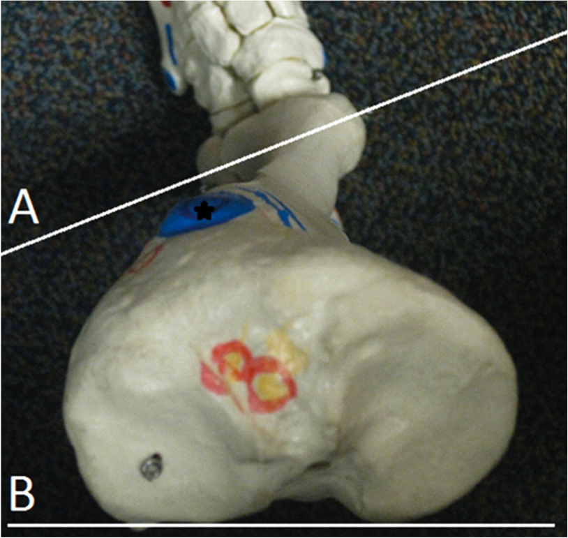
The final chapter of developmental versions involves the femur. The degree of version is the angle between an imaginary line drawn through the condyles of the femur and an imaginary line drawn through the head and neck of the femur. This is often referred to as the femoral neck angle or FNA.

Beginning about the 3rd month of embryological development (Lanz and Mayet 1953) and reaches about 40 degrees (with an average of 30-60 degrees) at birth. It then decreases 25-30 degrees by adulthood to 8-20 degrees with males being at the lower and females at the upper end of the range.

The FNA angle, therefore, diminishes about 1.5 degrees a year until about 15 years of age. Femoral neck anteversion angle is typically symmetrical from the left side to the right side.

What causes torsion in the first place? By the sixth month in utero, the lumbar spine and hips of the fetus are fully flexed, so perhaps it is positional. Other sources say it coincides with the degree of osteogenesis. There is a growing consensus that muscular forces are responsible, particularly the iliopsoas  or possibly the medial and lateral hip rotators.

Additional changes can occur after birth, particularly with sitting postures. “W” sitting or “cross legged” sitting have been associated with altering the available range of motion and thus the FNA, with the range increased in the direction the hip was held in; W sitting causing increased internal rotation and antetorsion and cross legged causing external rotation and retro torsion.

As discussed previously, there are at least 3 reasons we need to understand torsions and versions, They can alter the progression angle of gait, they usually affect the available ranges of motion of the limb and they can alter the coronal plane orientation of the limb.

1. fermoral torsions often alter the progression angle of gait.  In femoral antetorsion torsion, the knees often face inward, resulting in an intoed gait and a decreased progression angle of the foot. This can be differentiated from internal tibial torsion (ITT) by looking at the tibia and studying the position of the tibial tuberosity with respect to the foot, particularly the 2nd metatarsal. In ITT, the foot points inward while the tibial tuberosity points straight ahead. In an individual with no torsion, the tibial tuberosity lines up with the 2nd metatarsal. If the tibial tuerosity and 2nd met are lined up,  and the knees still point inward, the individual probably has femoral ante torsion. Remember that a decreased progression angle is often associated with a decreased step width whereas an increased angle is often associated with an increased step width. See the person with external tibial torsion in the above picture?

2. Femoral torsions affect available ranges of motion of the limb. We remember that the thigh leg needs to internally rotate the requisite 4-6 degrees from initial contact to midstance (most folks have 40 degrees) If it is already fully internally rotated (as it may be with femoral retro torsion), that range of motion must be created or compensated for elsewhere. This, much like internal tibial torsion, can result in external rotation of the affected lower limb to create the range of motion needed.

Femoral retro torsion results in less internal rotation of the limb, and increased external rotation.

Femoral ante torsion results in less external rotation of the limb, and increased internal rotation.

3. femoral torsions usually do not effect the coronal plane orientation of the lower limb, since the “spin” is in the transverse or horizontal plane.

The take home message here about femoral torsions is that no matter what the cause:

  •  FNA values that exist one to two standard deviations outside the range are considered “torsions”

  • Decreased values (ie, less than 8 degrees) are called “retro torsion” and increased values (greater than 20 degrees) are called “ante torsion”
  • Retro torsion causes a limitation of available internal rotation of the hip and an increase in external rotation

  • Ante torsion causes an increase in available internal rotation  of the hip and decrease in external rotation
  • Femoral ante torsion will be perpetuated by “W” sitting (sitting on knees with the feet outside the thighs, promoting internal rotation of the femur)

  • Femoral antetorsion will be perpetuated by sitting cross legged, which forces the thigh into external rotation.


Stay tuned for a case tomorrow to test your learning over the last few weeks.


We remain: Bald, good looking and intelligent…The Gait Guys


All material copyright 2013 The Gait Guys/ The Homunculus Group. All rights reserved.  Please ask to use our stuff!

Step width alters iliotibial band strain during running.

More substantiation that “the cross over gait” is a pathologic process.

Did you get to hear podcast #23 yet ?  Here is the link (iTunes).  In podcast #23 we talked at length about the effects of step width in runners.  Reducing ones step width will result in a progression into what we have been referring to for years as “the cross over gait”.  We have been reducing this phenomenon in our runners, and many walkers, for over a decade now to reduce many of the lower limb pathologic processes that ensue when the cross over is left unchecked and worse yet, strength and endurance is loaded upon the faulty pattern.  Everyone’s gait in this realm will differ because of pelvis width, femoral and tibial torsion, genu posturing (knee valgum, varum)  and foot structure and type. All of these factors must be taken into account when deciding upon the degree of step width correction.  Ultimately the goal in a perfect world would be to have the foot and knee stack pristinely under the centrated hip joint proper, but we all know that ideal biomechanics are the unicorn when it comes to humans. Anatomic variation is the known norm and this must not be forgotten, this was pounded into all of our heads in medical school.
As this article from the Nov 2012 J. of Sports Biomechanics clearly states, iliotibial band strain and strain rate is significantly greater in narrow based gait scenarios and that increasing step width during running, particularly in those who tend towards the lazier narrower step width, may be beneficial in not only the treatment but the prevention of future lateral hip and knee biomechanical syndromes such as IT band syndrome.  So, if you are a slave to your foam roller and need your IT band foam roller fix daily, you might want to look a little deeper at your biomechanics and make some changes.  Our videos here will be helpful to you and our writings on the Cross Over gait  and link here will be helpful as well.
In  summary, there is just so much more to good running form than just following the mantra “let my feet fall under my body mass and everything will be just fine”.  We wish it was this easy, but it is not. Unfortunately, too many of the sources on the internet are maintaining that good running form is mostly just that simple. Sadly, we find it our mission to bring the bitter tasting truth to the web when it comes to these things.  One just cannot ignore the factors of pelvis
width, femoral and tibial version and torsion, genu posturing (knee valgum, varum) and foot structure and foot type (and we mean so much more than are you a pronator or supinator).  These factors will alter lower limb biomechanics and may drive even the runner with heightened awareness of foot strike and running form into less than optimal foot strike positioning and loading response. Furthermore, one needs to be acutely aware that merely taking the cooked down under-toned postulation of this journal article, that being increasing step width will resolve their IT band problems, may not resolve their problem. In fact, without taking the issues of pelvis width, torsion, version, foot type and the like into account, making these changes could bring about more problems.  Seeking the advise of a knowledgeable physician in this complicated field of human locomotion is paramount to solve your chronic issues.
There is more to clean running than just a
midfoot-forefoot strike under the body mass, a good forward lean and high cadence. And we are here to bring those other issues to light, for the sake of every injured and frustrated runner.  Remember, uninjured does not always mean efficient. And efficient does not always mean uninjured.

Shawn and Ivo, The Gait Guys

_______________________________
Sports Biomech.
2012 Nov;11(4):464-72.Meardon SA, Campbell S, Derrick TR.

Step width alters iliotibial band strain during running.

Abstract
excerpted:

“Greater ITB strain and strain rate were found in the narrower step width condition (p < 0.001, p = 0.040). ITB strain was significantly (p < 0.001) greater in the narrow condition than the preferred and wide conditions and it was greater in the preferred condition than the wide condition. ITB strain rate was significantly greater in the narrow condition than the wide condition (p = 0.020). Polynomial contrasts revealed a linear increase in both ITB strain and strain rate with decreasing step width. We conclude that relatively small decreases in step width can substantially increase ITB strain as well as strain rates. Increasing step width during running, especially in persons whose running style is characterized by a narrow step width, may be beneficial in the treatment and prevention of running-related ITB syndrome.”

Podcast #24: Chronic achilles issues, beer recovery drink and case studies.

podcast link: http://thegaitguys.libsyn.com/pod-24-the-chronic-achilles-beer-cases

iTunes link: https://itunes.apple.com/us/podcast/the-gait-guys-podcast/id559864138

Show notes:

Gait Guys online store:

http://store.payloadz.com/results/results.aspx?m=80204

Today’s show notes:

1. J Trauma Acute Care Surg. 2013 Mar;74(3):946-7. doi: 10.1097/TA.0b013e31828272ad.
Achilles’ death: Anatomical considerations regarding the most famous trauma of the Trojan War.
2. J Foot Ankle Surg. 2001 May-Jun;40(3):132-6.Saxena A, Bareither D.

Magnetic resonance and cadaveric findings of the “watershed band” of the achilles tendon.
3. http://www.sciencedaily.com/releases/2013/02/130212112019.htm


Fallout from Nuclear Testing Shows That the Achilles Tendon Can’t Heal Itself
4. K. M. Heinemeier, P. Schjerling, J. Heinemeier, S. P. Magnusson, M. Kjaer. Lack of tissue renewal in human adult Achilles tendon is revealed by nuclear bomb 14C. The FASEB Journal, 2013; DOI: 10.1096/fj.12-225599

5.  http://www.washingtontimes.com/news/2013/feb/10/scientists-suggest-beer-after-workout/#.USRSIq-QMnw.facebook

Scientists suggest beer after a workout
6. Sports Biomech. 2012 Nov;11(4):464-72.

Step width alters iliotibial band strain during running.
7. _http://skorarunning.com/confessions-of-an-overpronator

Over-Pronation

8. http://www.championseverywhere.com/why-gait-analysis-doesnt-work-future-of-the-shoe-industry

Why gait analysis doesn’t work (future of the shoe industry

tumblr_min5irxcLp1qhko2so1_1280.jpg
tumblr_min5irxcLp1qhko2so2_1280.jpg

Yet another IFGEC certified individual, and our 1st in Asia! Congrats, Andy! Here’s what he has to say:

My name is Andy Neo aka Dee. I work as the regional senior tech rep for a distributor shoe company managing Singapore, Malaysia, Thailand & Indonesia. Additionally I graduated with a Bachelor degree in Sports & Outdoor from Monash University and an avid distance runner.   

I’m a keen learner about exercise physiology & human movement but there is no course for shoe fitting since I started working in the footwear industry few years back. A lot of knowledge was self learnt through trial & error because there are no industrial standards across the running shoes market. Fortunately I happened to tumble The Gait Guy’s blog & Facebook and was a keen follower of their regular posts. I was overjoyed when Dr Ivo & Dr Shawn in 2012 announced the opportunity to participate the National Shoe Fit Certification Examination online especially for me coming from another continent. The extensive 3hr lecture did helps to bridge the gap between health science, sports science & footwear education (seriously lacking in modern shoe industry). The lecture video was downloaded and studied at my own pace was really beneficial for working adults like myself. My advice to future IFGEC candidates is to have a good read up of human anatomy, shoe anatomy and human biomechanics for better understanding because the extensive 3hours lecture use technical terminology that can be jargon but definitely worthy information.

The quality of  examination questions required critical thinking & hence raise the standard of the certificate. Passing the IFGEC examination would elevate my profession so now I can better impart the knowledge to my retail staffs across 4 countries for a holistic shoe-fit service.

I recommend representative from performance footwear company & medical healthcare professions to spend quality time to sign up for this level 1 course because the contents will narrow the gap between health science & shoe industry.”


For more information on IFGEC certification, please email us at thegaitguys@gmail.com

http://thegaitguys.tumblr.com/post/21713480315/the-chef-another-abnormal-gait-pattern-in
Last night we caught a DVR&rsquo;d season show of Bourdain&rsquo;s new show &ldquo;the Layover&rdquo; on the Travel Channel.  Great new show, we love his diatri…

http://thegaitguys.tumblr.com/post/21713480315/the-chef-another-abnormal-gait-pattern-in

Last night we caught a DVR’d season show of Bourdain’s new show “the Layover” on the Travel Channel.  Great new show, we love his diatribes, rants and command of the English language. It reminded us of this post we did last year where we looked in depth at his unique gait flaw, the circumducting foot.

Join us again for this great gait dissection, see the link at the top. Even if you have been with us for over a year and still somewhat remember this gait pattern and our explanation of it, you will likely pick up another layer of  understanding after our last year of teaching  here on The Gait Guys blog.

Shawn and Ivo, The Gait Guys

tumblr_miivgh6NpC1qhko2so1_1280.jpg
tumblr_miivgh6NpC1qhko2so2_1280.png

Yes, we are all twisted. Part 3 continued.

If you missed yesterdays post, this one will make more sense if you go back and read it.Today we talk about compensations for tibial torsions.

As discussed in previous posts, there are at least 3 reasons we need to understand  tibial torsions and versions:

1. They will often alter the progression angle of gait.  In internal tibial torsion, there will often be a decreased progression angle of the foot and with external, an increased angle of progression. A decreased progression angle is often associated with a decreased step width whereas an increased angle is often associated with an increased step width.

2. They affect available ranges of motion (ROM) of the limb. We remember that the lower leg needs to internally rotate the requisite 4-6 degrees from initial contact to midstance:

ROM changes that may occur with internal tibial torsion

  • If it is already fully internally rotated (as it may be with internal tibial torsion), that range of motion must be created or compensated for elsewhere.
  • This can result in external rotation of the affected lower limb to create the range of motion neede
  • Circumduction of the lower limb, because the foot is already in a supinated posture, and the decreased range of motion of the foot needs to be compensated for.
  • A shortened step length, due to increased compressive forces at the medial knee
  • And alteration of vertical and medial lateral ground reactive forces
  • A rolling off the lateral aspect of the foot, due to it being in a more supinated posture

ROM changes that may occur with external tibial torsion          

  • external tibial torsion often results in the increased midfoot pronation, through the deformity, because more range of motion is possible both at the hip and foot at the subtalar joint

3. They often can effect the coronal plane orientation of the lower limb.

In internal tibial torsion, due to the foot being more rigid and the deformity often being accompanied by increased tibial varum, the knee often falls outside the plane of the foot (rather than being “stacked”), resulting in a decreased step width and often a cross over gait pattern (click here for more info on crossover)

In external tibial torsion, the foot is often more pliable. This often results in an increased step width and well as the knee falling inside (or medially) to the plane of the foot. Because of the increased hip and foot ranges of motion available,  the foot is not an adequate lever, shortening step length and sometimes requiring increased pelvic motion to “get around” the stance phase leg.

Whew! This stuff can be tough, Thanks for hanging in there! Next stop: Femoral Torsions and Versions!

Ivo and Shawn; your torsioned friends : )


All material copyright 2013 The Gait Guys/ The Homunculus Group. All rights reserved.  Ask before you lift our stuff, Lee is watching……

tumblr_mic2p8EQnN1qhko2so1_1280.jpg
tumblr_mic2p8EQnN1qhko2so2_1280.png
tumblr_mic2p8EQnN1qhko2so3_1280.gif

Yes, we are all twisted; Part 3


In the last 2 posts we discussed the differences between torsions and versions, as well as talar version and torsion, 1 of the 3 major versional events that occur during normal development (missed out? Click here and here to re read them).

In this post we discuss tibial versions and torsions.

The tibia and femur are more prone to torsional defects, as they are longer lamellar (layered) bones as opposed to the cancellous bone that makes up the talus. These often present as an “in toeing” or “out toeing” of the foot with respect to the leg; changing the progression angle of gait (click here for more on progression angles).

Tibial versions and torsions can be measured by the “thigh foot angle” (the angulation of the foot to the thigh with the leg bent 90 degrees: above right) or the “transmalleolar angle” (the angle that a line drawn between the medial and lateral malleoli of the ankle makes with the tibial plateau: above left). 

At a gestational age of 5 months, the fetus has approximately 20° of internal tibial torsion. As the fetus matures, The tibia then rotates externally, and most newborns have an average of 0- 4° of internal tibial torsion. At birth, there should be little to no torsion of the tibia; the proximal and distal portions of the bone have little angular difference (see above: top). Postnatally, the tibia should twist outward (externally) a total of 15 degrees until adult values are reached between ages 8 and 10 years of 23° of external tibial torsion (range, 0° to 40°). 

Sometimes the rotation at birth is excessive. This is called a torsion. Five in 10,000 children born will have rotational deformities of the legs. The most common cause is position and pressure (on the lower legs) in the uterus (an unstretched uterus in a first pregnancy causes greater pressuremaking the first-born child more prone to rotational deformities. Growth of the  unborn child accelerates during the last 10 weeks and the compression from the uterus thus increases. As you would guess, premature infants have less rotational deformities than full-term infants. This is probably due to decreased pressure in the uterus. Twins take up more space in the uterus and are more likely to have rotational deformities. 

Of interesting note, there is a 2:1 preponderance of left sided deformities believed to be due to most babies being carried on their backs on the left side of the mother in utero, causing the left leg to overlie the right in an externally rotated and abducted position.

Normal ranges of versions and torsions are highly variable (see chart above: right). Ranges less than the values are considered internal tibial torsion and greater external tibial torsion.

Internal tibial torsion (ITT) usually corrects 1 to 2 years after physiological bowing of the tibia (ie tibial varum) resolves. External tibial torsion (TT) is less common in infancy than ITT but is more likely to persist in later childhood and NOT resolve with growth because the natural progression of development is toward increasing external torsion.

Males and females seem to be affected equally, with about two thirds of patients are affected bilaterally and the differences in normal tibial version values are often expected to be cultural, lifestyle and posture related.

 The ability to compensate for a tibial torsion depends on the amount of inversion and eversion present in the foot and on the amount of rotation possible at the hip. Internal torsion causes the foot to adduct, and the patient tries to compensate by everting the foot and/or by externally rotating at the hip. Similarly, persons with external tibial torsion invert at the foot and internally rotate at the hip. Both can decrease walking agility and speed if severe. With an external tibial torsion deformity of 30 degrees , the capacities of soleus, posterior gluteus medius, and gluteus maximus to extend both the hip and knee were all reduced by over 10%.

Well, that was probably more than you wanted to know about tibial torsions, and we could go on for many more pages and perhaps cure any insomnia you may have. Take a while to digest this, as it is important to gait, shoe selection, and rehabilitation. Torsions are an acquired taste and we hope we have whetted your appetite! Tomorrow we talk about compensations!


Ivo and Shawn; two twisted guys!


All material copyright 2013 The Gait Guys/ The Homunculus Group. All rights reserved.  Ask before you lift our stuff, Lee is watching……

Podcast #23. Neurology of walking babies, dialogues on step width for runners and so much more !

Syndication link:

http://thegaitguys.libsyn.com/podcast-23-walking-babies-step-width-cross-over-running

iTunes link:

https://itunes.apple.com/us/podcast/the-gait-guys-podcast/id559864138

Podcast #23. Neurology of walking babies, dialogues on step width for runners and so much more !

1- Newborn babies walk the walk | Body & Brain
http://www.sciencenews.org/view/generic/id/348262/description/Newborn_babies_walk_the_walk

2- 3D printing with stem cells could lead to printable organs - CNET Mobile
http://m.cnet.com/news/3d-printing-with-stem-cells-could-lead-to-printable-organs/57567789

3- our payloadz e-file download site.  http://store.payloadz.com/results/results.aspx?m=80204

4- www.onlineCE.com  

Great TeleSeminar Wed Feb 20th, 2013 8:00 PM Eastern Time Chiropractic TeleSeminar Biomechanics 302 Location: 1 hr by telephone Instructor: Waerlop/Allen, DC Price: 19.00

5- J Biomech. 2004 Jun;37(6):935-8.Owings TM, Grabiner MD.  Step width variability … .

Brach JS.    J Neuroeng Rehabil. 2005 Jul 26;2:21.  Step width variability … .

Sports Biomech. 2012 Nov;11(4):464-72.  IT Band strain and step width … .

6. Rethinking Ice Baths And Ibuprofen pulse.me/s/isg3t Inflammation IS part of the healing process!

7. Bringing the Foot Back To Life: Restoring the Extensor Hallucis Brevis Muscle.

http://youtu.be/1iZg_e4veWk