Needling Myofascial Meridians?

15079418828_c1a437b173_b.jpg

Beyond the Trigger Point...

Many clinicians needle. We are taught to dry needle trigger points and to needle the segmental innervation of the muscle involved. But should we do more? I think so, and here is one paper on incorporating needling myofascial meridians along with trigger points that supports that notion (1).

Since most of us treat patients that are ambulatory, we should be thinking of how a patient moves, especially through the gait cycle. Think of the kinetic chain in what I like to call “reverse engineering”, that is, from the ground up, rather from the torso down, in a closed chain fashion. This will profoundly effect the way you look at muscle function, for example: thinking of the vastus lateralis as a medial rotator of the thigh (yes, you read that right; think about it and try and get your head wrapped around it), or of the peroneus longus as an abductor of the lower leg and external rotator (assisting supination) during the latter half of the gait cycle. Reverse engineering gives you a whole new outlook on locomotion and human movement.

Tom Myers was insightful enough to write a great text talking about myofascial meridians or “lines of tissue stress ” describing the fascial connections of muscles utilized in a chain during movement (2). This built upon the original work of Valdimir Janda and his concepts of “loops and slings” (3), as well as the work of Paoletti (4) and Vleeming (5). 

In neurology, we have the conjoint concepts of temporal and spacial summation that can lead to some action on the part of the nervous system. They describe 2 ways that receptors or neurons can reach threshold and fire an action potential (6) .

Temporal summation is when a receptor or neuron is stimulated repetetively over time, with each potential bulding upon the previous, making the stimulus effectively larger and larger. If you were in a movie theater and the person seated behind you kept hitting the back of your seat repetitively (temporal summation), it would only be a matter of time before you turned around and said some thing to them(ie, you reached threshold).

Spacial summation is when a receptor or neuron is stimulated at multiple locations over time, with the potentials building and bringing the receptor or neuron closer to threshold.  Taking the same scenario as before, if many people began hitting your chair from multiple directions (spacial summation), it would be only a matter of time before you said something (ie: reached threshold).

These two things can work together as well, usually eliciting a result much faster, since the receptor or neuron is being hit multiple times from multiple directions and it can usually reach threshold faster.

Since one of our goals in needling is not only to reduce or eliminate the trigger point, but also to reduce pain and increase function, wouldn’t it make sense to take advantage of as much neurology as possible? How about more real estate (spacial summation) in a reasonable time frame from point to point (temporal summation)?

Needling appears to cause pain modulation, as well as many of its other effects,  through both peripheral and central mechanisms (7,8). Having our therapy stimulate more of these mechanisms should theoretically make our therapy more effective and improve outcomes. So, more needles may be a good thing, no? 

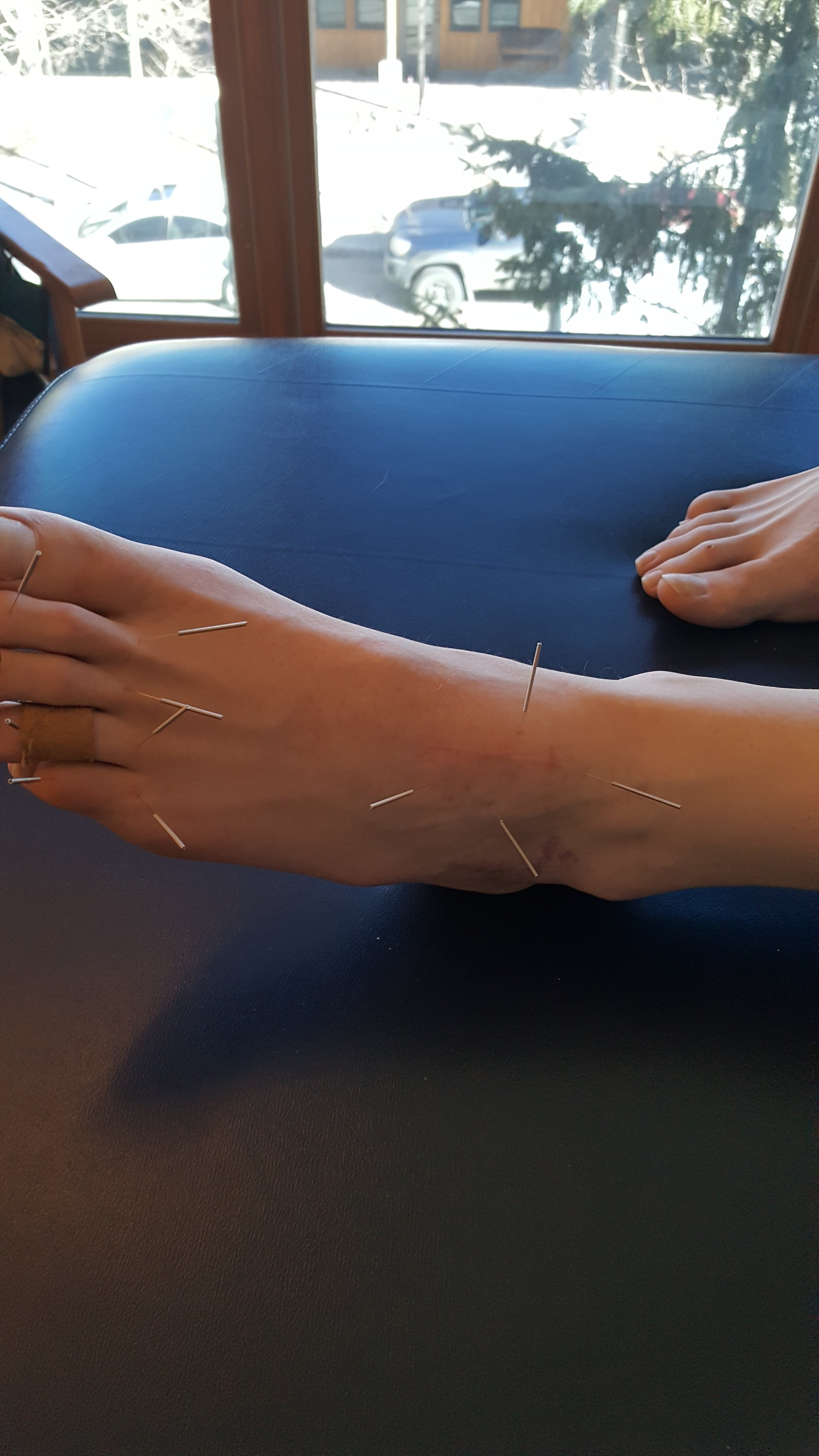
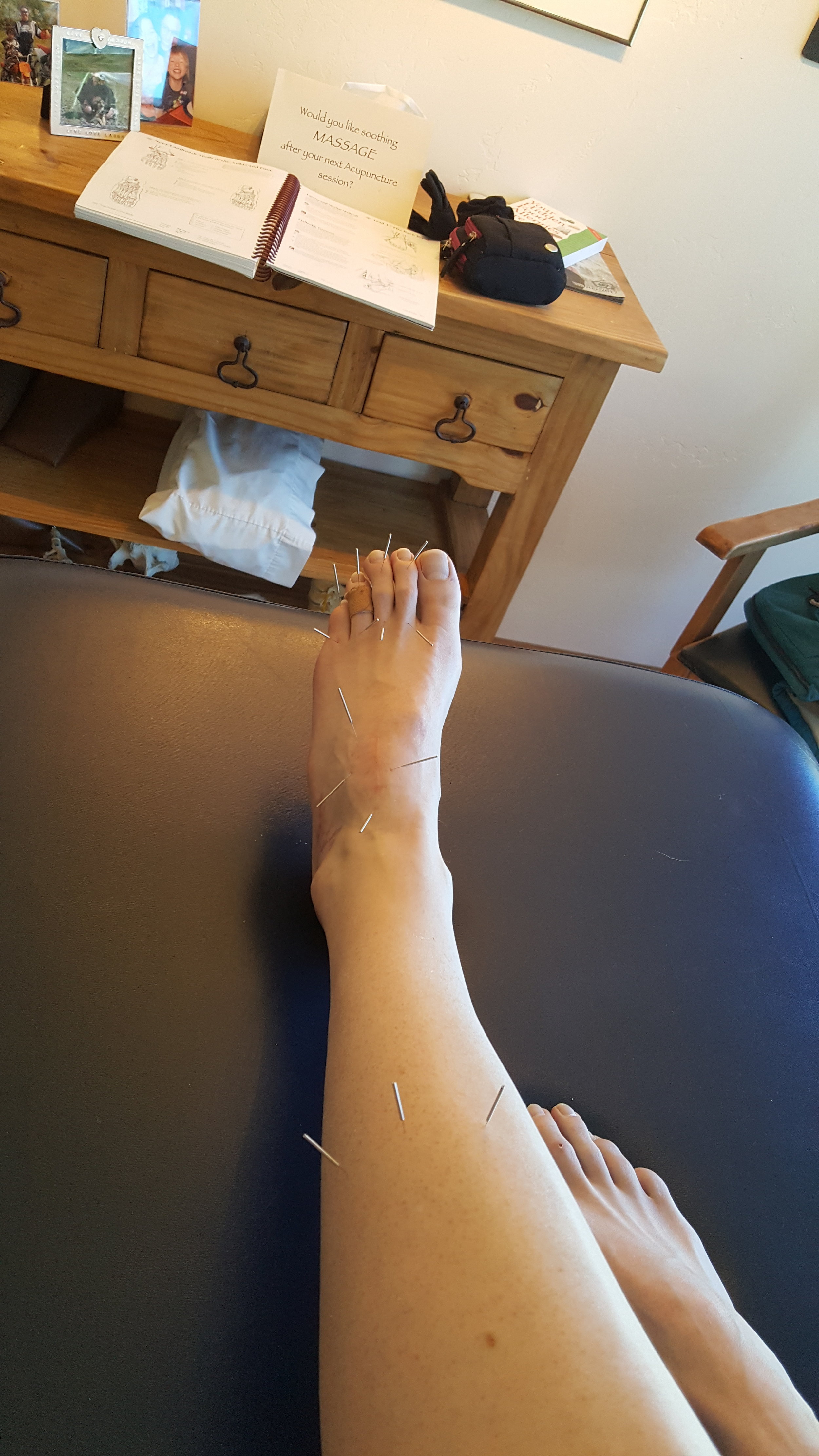
Getting back to the paper (1), they needled tender points (satellite trigger points?) along the lower portions of the “superficial back line” or “SBL”, along with points on the foot for plantar fascitis. The SBL contains plantar fascia, Achilles tendon, gastrocnemius,hamstrings, sacrotuberous ligament, and erector spinae. It continues to the suboccipital muscles and ends at the suboccipital muscles, galea aponeurotica of the skull and ultimately the frontalis muscle (2). They could have incorporated more, and perhaps had even better results, as the upper cervial spine contains one of the highest densities of mechanoreceptors in the body (9, 10), and afferent information from the upper 4 cervical neuromeres feed directly into the flocculonodular lobe of the cerebellum (11, 12).

So, how about incorporating myofascial meridians into your needling toolbox? The next time you see someone with a problem area, think about the kinetic chain that gets you there, starting from the ground up, and incorporate THAT into your treatment protocol. 


references:

1. Akhbari B, Salavati M, Ezzati K,  Mohammadi Rad S: The Use of Dry Needling and Myofascial Meridians in a Case of Plantar Fasciitis Journal of Chiropractic Medicine (2014) 13, 4348

2. Myers TW. Anatomy trains: myofascial meridians for manual and movement therapists. 2nd ed. Philadelphia: Churchill Livingstone; 2009.

3. Janda V, Vavrova M, Hervenova A, et al. Sensory motor stimulation. In: Liebenson C. ed Rehabilitation of the spine: a practitioners manual. 2nd edn. Lippincott Williams & Wilkins, 2006.

4. Paoletti S. The fasciae: anatomy, dysfunction & treatment. Eastland Press; 2006.

5. Vleeming A, Snijders C, Stoeckart R, Mens J. The role of the sacroiliac joins in coupling between spine, pelvis, legs and arms. In: Vleeming A, et al, editor. Movement, stability and low back pain. Churchill Livingstone; 1997. p. 5371

6. Levin & Luders (2000). Comprehensive Clinical Neurophysiology. New York: W.B. Saunders Company.

7. Dommerholt j Dry needling — peripheral and central considerations Journal of Manual and Manipulative Therapy 2011 VOL. 19 NO. 4 223-237

8.  Li-Wei Chou,  Mu-Jung Kao, Jaung-Geng Lin  ProbableMechanisms of Needling Therapies for Myofascial Pain Control Evidence-Based Complementary and Alternative Medicine Volume 2012, Article ID 705327, 11 pages doi:10.1155/2012/705327

9. Kulkarni V1, Chandy MJ, Babu KS  Quantitative study of muscle spindles in suboccipital muscles of human foetuses. Neurol India. 2001 Dec;49(4):355-9

10. Bogduk N Cervicogenic headache: anatomic basis and pathophysiologic mechanisms. Curr Pain Headache Rep. 2001 Aug;5(4):382-6.

11.   Luan H1, Gdowski MJ, Newlands SD, Gdowski GT  Convergence of vestibular and neck proprioceptive sensory signals in the cerebellar interpositus. J Neurosci. 2013 Jan 16;33(3):1198-210a. doi: 10.1523/JNEUROSCI.3460-12.2013.

12.  Seaman D Winterstein  Dysafferentation:   A Novel Term to Describe the Neuropathophysiological Effects of  Joint Complex Dysfunction. A Look at Likely Mechanisms of Symptom Generation  J Manipulative Physiol Ther 1998 (May);   21 (4):   267-280

Something a little different for a change: Case Management of a Post Surgical Foot

In this series, we will follow the progress of a post surgical, post rehab foot. These are the actual case notes and you can follow our thought process as we move along. 

History:

JM presented with left-sided content foot pain. On July 24 she broke her left navicular and cuboid (pretty unusual, as these fractures are rare. Navicular fractures are usually stress fractures (1), occurring in about .6% of fractures in one study (2).  Cuboid fractures are also rare and occur in less than 1.8 per 100,000 (3) ) She also tore the reticular ligaments. this happened when she fell down the stairs, inverting and plantar flexing the foot.

She has had extensive physical therapy as well as plate fixation of the navicular but is still having constant discomfort; she feels a pinching and shock like sensation in the right arch, particularly when loading the foot (whenever you hear about a "shock like pain, begin thinking about nerve related pain). She has been on gabapentin in the past which helped but she stopped it 3 weeks ago with no regression of her symptoms. She feels frustrated.

She was in physical therapy until the end of December. She has continued with exercises consisting of plantar flexion/toe raises, mobilization and inversion/eversion, squats/lunges as well as massage. She has improved but not completely better. She is able to hike 4-5 miles with little pain (boy, those Colorado women are tough!). The foot generally feels better with non weight bearing and rest as well as avoiding impact. The foot feels stiff in the morning and  she limps for the first 10 minutes after getting out of bed. Most recently she has had x-rays at VSO with Dr. X.

What did we find?

There was swelling noted over the extensor digitorum brevis with significant weakness of it as well as the extensor longus. No sensory deficits, reflexes intact. She had an anatomically short left short leg which appeared to be functional. There was a scar visible over the dorsum of the foot approximately 2 inches in length (see photos) and some discoloration lateral just anterior to the lateral malleolus. palpation along the medial plantar nerve revealed increased sensitivity below the navicular and into the medial heel. 

She has external tibial torsion bilaterally and limited eversion of the forefoot on the right. Her cuboid was moving appropriately but talonavicular articulation was not. She has adequate hip extension, 15+ degrees and ankle dorsiflexion bilaterally in excess of 15 degrees.

One leg standing with eyes open was less than 10 seconds. Loss of flexion and extension about the L SI joint. 

no x rays available for this visit

What we think was wrong:

Left foot pathomechanics (talonavicular) secondary to surgery and fracture. She has significant weakness of the short and long extensors as well as limited eversion and proprioceptive difficulties.

Discussion:

The talonavicular articulation is one of the key joints in mid foot pronation. Pathomechanics appear to be compromising the medial plantar nerve. This is exacerbated by her inability to fully evert the forefoot and pronate through the mid and forefoot (pronation is dorsiflexion, eversion and abduction). 

What We did:

We manipulated the sacroiliac joint and metatarsophalangeal articulations. We held off on any mobilization of the foot until we see her x-rays, hopefully available next visit.

We treated with neuromuscular acupuncture at the origin/insertion of the long extensors as well about the short extensor mass where the swelling was located. She was given the tripod standing, lift spread reach, toes up walking, and tiptoe waiting exercises to perform 2-3 times daily. She is scheduled for followup next week with x-ray review.


1. http://emedicine.medscape.com/article/85973-overview

2. http://journals.sagepub.com/doi/pdf/10.1177/2473011416S00299

3. http://www.uptodate.com/contents/cuboid-and-cuneiform-fractures

Dry Needling and Muscle Activation Patterns

A nice study looking at how sequential muscle activation patterns can change with dry needling. Think about the applications for gait?

"Removing LTrPs changes the order of muscle recruitment to a more sequential, stable pattern that is not significantly different to that displayed by the control group prior to fatiguing exercise. This suggests that removing LTrPs may allow subjects to better cope with the effects of fatigue, as evidenced by the reduced variability in activation times and the reduced co-activation of the muscles investigated. "

FREE FULL TEXT here: https://isbweb.org/images/conf/2003/longAbstracts/LUCAS_198-208_SB_LONGE.pdf

Dry Needling and Myofascial Pain

Regardless of the mechanism, dry needling and ischemic compression both seem to reduce myofascial pain. How about some more studies looking at muscle function and activation patterns?

"This study compared these treatment techniques to one another using the Neck Disability Index (NDI), a numeric rating scale (NRS), pressure pain threshold and muscle characteristics. 42 female patients with myofascial neck pain were randomly assigned to a treatment group and the 4 most painful MTrPs were treated using DN or MPT. No difference was found between the two techniques on the short and long term. Both techniques showed an improvement in NDI on the short and long term. "

Dry needling or manual pressure in myofascial pain? - Anatomy & Physiotherapy

The aim of this study was to compare dry needling to manual pressure in patients with myofascial pain.

ANATOMY-PHYSIOTHERAPY.COM|BY <A HREF="/AUTHORLIST/3:JOANNA1988" TITLE="VIEW ALL ARTICLES FROM JOANNA TUYNMAN">JOANNA TUYNMAN</A>


Those MultifidiThe multifidi are important proprioceptive sentinels for the low back, as well as the rest of the body, for virtually every activity you do weight bearing, including gait. They are implicated in many instances of low back pain, especi…

Those Multifidi

The multifidi are important proprioceptive sentinels for the low back, as well as the rest of the body, for virtually every activity you do weight bearing, including gait. They are implicated in many instances of low back pain, especially folks with flexion or extension intolerance, since their fiber orientation and thus mechanical advantage (or disadvantage) is dependent upon whether or not you are maintaining a normal lumbar lordosis.

Modalities which boost their function are an excellent adjunct to the rehabilitation process. Since they are not under volitional control (go ahead, try and contract your L2/L3 multifidus), they are innervated by the vestibulospinal tract and we must use proprioceptive work to engage them. Dry Needling is one modality that can help them to become functional again.

RESULTS and CONCLUSION:
“Significant difference was found in the percentage of change of muscle activation post needling between groups on the right side at level L4-5. A slight increase in the percentage of muscle activity, post procedure was observed in the dry needling group compared with the control group, although not significant in other segments examined. An improvement of back muscle function following dry needling procedure in healthy individuals was found. This implies that dry needling might stimulate motor nerve fibers and as such increase muscle activity.”

see also our post here.

J Back Musculoskelet Rehabil. 2015 Sep 6. [Epub ahead of print]
The immediate effect of dry needling on multifidus muscles’ function in healthy individuals. Dar G1,2, Hicks GE3.

tumblr_o9vc4xvrkE1qhko2so1_400.jpg
tumblr_o9vc4xvrkE1qhko2so2_400.jpg

One point and 1 treatment can profoundly influence gait

When talking about the lower extremity and gait (as I have been know to do at more that one seminar), I often talk about the “reverse engineering” principle. This is looking at a muscle or muscle group from a “ground up” perspective, as it would be functioning during the gait cycle. This, along with knowing when a muscle should be firing in the gait cycle, can provide clues to what may be going on and how you may be able to help.

When discussing the quads, we often employ this principle. It can be a little difficult to think of the vastus medialis as a lateral rotator of the thigh and the rectus femoris as a flexor (anterior nutator) of the pelvis, but if you put your foot on the ground and think about it, you will see what I mean.

The VMO is often implicated in patello femoral syndromes but cannot be selectively activated. The ratio between vastus medialis and vastus lateralis does seem to be alterable and perhaps is a siginificant factor.

How about if we look at the vastus lateralis instead?

The vastus lateralis is the largest and most powerful portion of the quadriceps. One paper reports that the muscle volume of the the vastus lateralis was 674 cm3 followed by the vastus intermedius at 580 cm3, vastus medialis 461 cm3 and lowest in the rectus femoris 339 cm3.  This makes the vastus lateralis is twice the volume of the rectus femoris!

Studies of muscle fiber orientation show that VL force component is directed approximately 12-15° laterally with respect to the longitudinal axis of the femoral shaft. This would mean it has a tremendous mechanical advantage and could (should?) pull the patella directly laterally compared to the VMO force, whose component is directed approximately 55 ° medially.   The muscle “balance” between the VMO and the VL, along with the periarticular soft tissue structures acting on the patella, is considered major component in the control of normal patellar alignment and function. The VL is often considered to be the “overactive” one by many clinicians, particularly in cases of patellofemoral dysfunction. It turns out that from an EMG standpoint, they may be correct. 

The vastus lateralis arises posteriorly from the femur along the linea aspera and circumnavigates the thigh in a counterclockwise fashion to attach laterally to the patellar tendon.   Because of its size and fiber orientation, it would stand to reason that needling it would have more cortical representation than say the vastus medialis.

There is an interesting paper where they needled a single acupuncture point: Stomach 34. For those who haven’t studied acupuncture (or don’t remember) this point is located on the thigh, in a small depression about 2.5 inches (63 mm for the metric folks) lateral to and above lateral border of the patella. In other words, it is in the vastus lateralis (see above).

The results showed statistically significant improvement in velocity, cadence, stride length, cycle time, step time and single/double leg support after treatment. The effect was small, but positive.

Think about where the trigger points are for this muscle (see above) ; fairly close to this point, sometimes (depending on the trigger point), even directly over this point. Needling has many effects on muscle and its trigger points and we like to think that needling “normalizes” function of a muscle; perhaps it influences the apparent “dominance” of this muscle and allows the patella to track more medially?

So, in this popultion of patients of elderly individuals, 1 acupuncture (needling) treatment  had a positive influence on their gait. Perhaps if the folks in the knee study were treated a few more times, we would have seen a change. Imagine what could have happened if aditional treatment modalities, like exercise, proprioceptive work and gait retraining were added! 

What a great, cost effective alternative or addition to your rehabilitation this could be. Consider adding this modality (and point!) to your current clinical toolbox, not only for older patients but for any patients that may have a gait abnormality.


https://tmblr.co/ZrRYjx24ecKUx

Boucher JP, King MA, Lefebvre R, Pépin A. “Quadriceps femoris muscle activity in patellofemoral pain syndrome.” Am J Sports Med. 1992 Sep-Oct;20(5):527-32. Web. 17 Nov 2012.

Souza DR, Gross MT. “Comparison of vastus medialis obliquus: vastus lateralis muscle integrated electromyographic ratios between healthy subjects and patients with patellofemoral pain.” Phys Ther. 1991 Apr;71(4):310-6. Web. 25 Nov 2012.

Cowan SM, Bennell KL, Crossley KM, Hodges PW, McConnell J. “Physical therapy alters recruitment of the vasti in patellofemoral pain syndrome.” Med Sci Sports Exerc. 2002 Dec;34(12):1879-85. Web. 26 Nov 2012.

Boling MC, Bolgla LA, Mattacola CG, Uhl TL, Hosey RG. “Outcomes of a weight-bearing rehabilitation program for patients diagnosed with patellofemoral pain syndrome.” Arch Phys Med Rehabil. 2006 Nov;87(11):1428-35

Kim, H. H., & Song, C. H. (2010). Effects of knee and foot position on EMGactivity and ratio of the vastus medialis oblique and vastus lateralis during squatexercise. Journal of Muscle and Joint Health, 17(2), 142-150.

Lam, P. L., & Ng, G. Y. (2001). Activation of the quadriceps muscle during semisquatting with different hip and knee positions in patients with anterior knee pain. American Journal of Physical Medicine & Rehabilitation, 80(11), 804-808.

Erskine, R. M., Jones, D. A., Maganaris, C. N., & Degens, H. (2009). In vivo specific tension of the human quadriceps muscle. European journal of applied physiology, 106(6), 827-838. [PubMed]

Grabiner MD: Current Issues in Biomechanics (9th ed). Champaign, Human Kinetics Publishers, 1993.

http://www.orthobullets.com/anatomy/10058/vastus-lateralis

Hauer K, Wendt I, Schwenk M, Rohr C, Oster P, Greten J. Stimulation of acupoint ST-34 acutely improves gait performance in geriatric patients during rehabilitation: A randomized controlled trial. Arch Phys Med Rehabil. 2011 Jan;92(1):7-14. doi: 10.1016/j.apmr.2010.09.023.

Peter Deadman, Mazin Al-Khafaji, Kevin Baker: A Manual of Acupuncture (2nd Edition) Journal of Chinese Medicine Esat Sussex, England 2007

Travell JG, Simons DG. Myofascial Pain and Dysfunction: The Trigger Point Manual: The Lower Extremities. Vol.2 . Baltimore, Md: Williams & Wilkins;1992

 http://www.medscape.org/viewarticle/521494_3

The Vasti

Do you treat runners? Do you treat folks with knee pain? Patellar tracking issues? Do you treat the quadriceps? Do you realize that the vastus lateralis, in closed chain, is actually an INTERNAL rotator of the thigh (not a typo), and many folks have a loss of internal rotation of the hip? Do you give them “IT band stretches” to perform?

In this short video, Dr Ivo demonstrates some needling techniques for the quads and offers some (entertaining) clinical commentary on the IT band. A definite view for those of you who have needling in their clinical tool box.

Dry Needling and Proprioception. What a great combination. Since dry needling and proprioception both have such profound effects on muscle tone, why not combine them to treat chronic ankle instability? We do all the time and here is a FREE FULL TEXT…

Dry Needling and Proprioception. What a great combination.

Since dry needling and proprioception both have such profound effects on muscle tone, why not combine them to treat chronic ankle instability? We do all the time and here is a FREE FULL TEXT article that ties the two together nicely!

And what better to muscle to use than the peroneii? These babies help control valgus/varus motions of the foot and influence plantar and dorsiflexion AND the longus descends the 1st ray. We call that a triple win!

“This study provides evidence that the inclusion of TrP-DN within the lateral peroneus muscle into a proprioceptive/strengthening exercise program resulted in better outcomes in pain and function 1 month after the end of the therapy in individuals with ankle instability. Our results may anticipate that the benefits of adding TrP-DN in the lateral peroneus muscle for the management of ankle instability are clinically relevant as large between-groups effect sizes were observed in all the outcomes.”

link to full text
http://www.ncbi.nlm.nih.gov/pmc/articles/PMC4430654/

photo from this past weekends Dry Needling Seminar: working on the dorsal interossei

Welcome to Monday and News You Can Use!

Any of your patients of clients taking anti inflammatories? Especially after a rehab session or dry needling/acupuncture? They may be thwarting the healing process. Excerpted from a recent lecture, Dr Ivo talks about how they can down regulate the healing process.