Rewind: Walking gait in a pencil skirt

Gait Pathomechanics: Walking in a Pencil Skirt.

We wrote this piece 5 years ago. We are updating it with a new disasterous video. Speed ahead to the 30 second mark to get to the good stuff. The Gait stuff.
No longer can there be adequate use of the obliquity of the pelvis and thus abdominals or contralateral leg swing to initiate supination and toe off and . . .

blog link: https://www.thegaitguys.com/…/gait-pathomechanics-walking-i…

https://www.youtube.com/watch?v=LSVzlRouRk4

The glutes are in fact great internal hip rotators, too. Open your mind.

The glutes as an internal rotator ? Yes, to understand squats and lunges and deep hip flexion activities one needs to understand that the glutes , which we typically perceive as hip extensors and external rotators, can also assist in internal rotation. Which, we explain in this 2 years old rewind blog post.
We discussed this at length in our online teleseminar last night. Join us for this and other deep gait and biomechanics topics every 3rd Wednesday of the month !

Screen Shot 2018-04-06 at 8.17.11 AM.png

I recently got a message from a colleague questioning as to how in the world, that when the hip is in flexion, the glutes and piriformis become internal rotators.  This is again another example of lack of functional anatomy knowledge.  It took me awhile to find a picture to help explain this, but I finally found one reasonable to do so. Many readers who are stuck on this concept are just too stuck on the anatomy as presented in the image to the right, neutral stance-like.  This article today will be all about internal and external moment arms, here, this lecture will help a little, it is on glute medius internal moment arms in stance phase however, so there is little carry over but it will at least get you understanding moment arms more clearly. 

We tend to just think of the glute max as a hip stabilizer and extensor, for the most part. It also decelerates flexion in terminal swing.  The glute medius is mostly thought of as a lateral hip stabilizer and abductor, either of the femur (open chain) or of the pelvis in stance position (closed chain), meaning zero degrees or neutral plus or minus the trivial degrees of engaged hip flexion and extension used in normal gait.

No one I know consciously trains the glutes as an internal rotator, but there are many actions where we need this function, such as in crawling and many high functioning activities such as martial arts grappling and kicking for example. Gymnasts should also know that the glutes are powerful internal hip rotators.  If you are doing quadruped crawling work you also need to know this as your client approaches 90 degrees of hip flexion. No one ever seems to check this critical gluteal function, at least I see it missed all the time from my referring doctors and therapists for unresolving hip pain cases. Patients with hip pain, anterior, lateral or posterior, with lack of internal hip rotation need the glutes checked just as much as the other known internal hip rotators we all seem to know (though some still do not understand how powerful the vastus lateralis is as an internal rotator, but again, those are folks who just have not spend the time in a mental 3D space looking at functional anatomy. I live mentally in that 3D space all day long when working with patients, you should too.) Let me be more clear, the anterior bundle, the iliac bundle of the glute max, is an internal rotator in flexion, the sacral and coccyxgeal divisions are not, they are external hip rotators in flexion. The gluteus medius and minimus are internal hip rotators closing in on 90 degrees hip flexion.  Hence, you must be able to tease out these divisions in your muscle testing, one cannot just test the glutes as external rotators or extensors, you are doing a really sloppy job if that is all you are doing. Nor should someone just train the glutes as hip stabilizers, external hip rotators and extensors (which is probably 90% of the trainers and coaches out there I might assume?). IF one knows the origin and insertions (see the blue and green arrows) and moves those points towards each other in a fashion of concentric contraction (purple arrows) one should be able to easily see that this will orient the femur to spin into internal rotation in the acetabulum (follow the arc of the black arrows). The same goes for eccentric contractions, it is the same game.  If you are doing DNS and crawling work, you should know this stuff cold gang. When you close chain the hip in sitting, or are moving from tall kneeling into flexed kneeling chops, performing high knees in sprint training,  or especially in crawling and climbing type actions, you must understand the mechanisms of internal rotation creation and stabilization -- if the glutes are not present and trained and useful in flexion, you are missing a chunk of something big. Amongst many other things, your client must be capable, stable, strong and skilled in moving from supine to quadruped all in one turning-over motion to teach how to stabilize the hip in the quadruped action and then progress into crawling.  This is a reflexive action learned in the early motor developmental phase of locomotion.  So take your client back through this motor pattern if they have some of the hip problems with internal rotation, it is a small piece of the gluteal puzzle.

I am sure this will show up in someone's seminar at some point, hopefully it is in many already, it has always been in my lectures when going down the rabbit hole of all things glutes. And to be fair, I haven't been to seminars in years as I get too frustrated, so this concept may be everywhere for all I know (lets hope).  But that is something I have to get over, I am sure I still have much to learn.  

To give credit where credit is due, which we always insist upon here at The Gait Guys, this was refreshed in my mind by Greg Lehman in a Facebook post forwarded to me by the inquiring doctor.   Link here  and from the article that spurred him to discuss it, an old article I read long ago just after completing my residency, the article is by Delp et al.  It is worth your time.  Thanks Greg for bringing this back into the dialogue, it is critical base knowledge everyone should already know. 

Variation of rotation moment arms with hip flexion.  Scott L. Delp,*, William E. Hess, David S. Hungerford, Lynne C. Jones  J. of Biomechanics 32, (1999)

-Dr. Shawn Allen, the other Gait Guy

link: https://www.thegaitguys.com/thedailyblog/2016/12/7/the-glutes-are-in-fact-great-internal-hip-rotators-too-open-your-mind

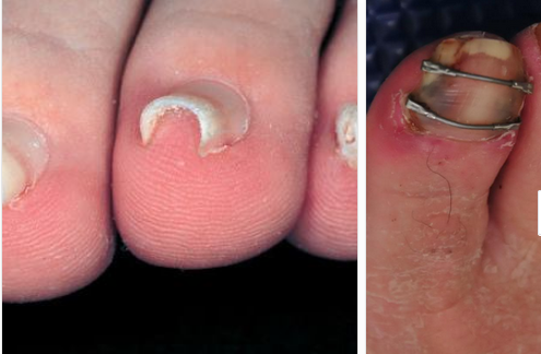
Pincer Toe nails: You've seen them; did you know what they were and how they got that way? Or, did you dismiss them?

Screen Shot 2018-04-06 at 8.13.23 AM.png

We think Hitomi’s hypothesis is correct. Here is why (this is paraphrased from our blog post on subungal hematomas and our revolutionary thinking on why they occur and it seems to fit well with pincer nail formation as well).

… when the skin is pulled at a differential rate over the distal phalange (from gripping of the toes rather than downward pressing through the toe pad) there will be a net lifting response of the nail from its bed as the skin is drawn forward of the backward drawn phalange (there is a NET movement of skin forward thus lifting the nail from its bedding). For an at-home example of this, put your hand AND fingers flat on a table top. Now activate JUST your distal long finger flexors so that only the tip of the fingers are in contact with the table top (there will be a small lifting of the fingers). There should be minimal flexion of the distal fingers at this point. Note the spreading and flattening of the nail. Now, without letting the finger tip-skin contact point move at all from the table, go ahead and increase your long flexor tone/pull fairly aggressively. You are in essence trying to pull the finger backward into flexion while leaving the skin pad in the same place on the table. Feel the pressure building under the distal tip of the finger nail as the skin is RELATIVELY drawn forward.] This is fat pad and skin being drawn forward (relative to the phalange bone being drawn backward) into the apex of the nail. Could this be magnifying the curvature of the nail and not offsetting the “automatic curving and shrinkage” function of the nail ? We think it is quite possible.

We have more to say on this topic, the above is just an excerpt of our blog post. More here, in the link below

The season to pathologize our feet is upon us. Toe extension matters.

Screen Shot 2018-04-06 at 8.05.18 AM.png

I blew out my flip flop,
Stepped on a pop top;
Cut my heel, had to cruise on back home.
But there's booze in the blender,
And soon it will render
That frozen concoction that helps me hang on. - Jimmy Buffett

I continue to see more and more people with inadequate toe extension. It is complicated. I see those who do not even have the awareness of toe extension, loss of strength of toe extension, loss of endurance of toe extension, loss of global range of toe extension (dorsiflexion at the MTP joint), more failure of long toe extensor (EHL) strength and more prominence of increased short toe extensor strength (EDB) and more frightening, a lack of disassociation of toe extension (MTP dorsiflexion) and ankle dorsiflexion. Many clients when asked to life their toes, will drive into ankle mortise dorsiflexion; ask them to just purely toe dorsiflex and the mental games begin, a wrinkled brow, intense concentration. If you cannot extended the toes sitting, how are you going to find them in swing phase of gait when balance, and other things, are more important?
Stand and lift your toes. The arch should go up, you have engaged the Windlass Mechanism, that winds up the plantar fascia and raised the arch. If you do not have competent, unconsciously competent, toe extension, your arch is not all that it can, and should, be. If you cannot raise your toes, thus raise the arch, thus plantarflex the first metatarsal, then in gait, when the foot is on the ground, you cannot properly position the sesamoids, properly get safe terminal ranges of hallux dorsiflexion at toe off, properly position the foot for loading and unloading, adequately achieve ankle dorsiflexion, adequately offer the hip a chance for ample hip extension, offer the glutes optimal chance to work in all phases to help control spin of the limb during loading and unloading, and the list goes on and on. I am sure I left much out there, this was written in a few minutes and unedited, just a short rant for the weekend. But if you have not championed toe extension, both in an unloaded and loaded foot (on the ground), achieved control of both long and short extensor muscles to the toes (and paired them well with the long and short toe flexors), disassociated toe extension from ankle dorsiflexion, and then figured out how to properly, timely, engage all these processes into your gait unconsciously, you are working on less of an optimal system than you should be. So, if your feet hurt, hips hurt, or a plethora of other problems that you are trying to fix with orthotics or other toys, maybe start with, "can you lift your toes?". It is a piece of the puzzle, trust me.
Or, you can just stay in your flip flops and perpetuate your toe flexion and wait for bad things to take root After all, tis the season soon !
Yes, toe extension in flip flops (we must flex our toes to keep them on) is as rare as a good multi-tasking man.

Shawn Allen, one of the gait guys.

Functional Ankle Instability and the Peroneals

tumblr_mgcawrDlDn1qhko2so1_400.jpg

Lots of links available here with today’s blog post. please make sure to take your time and check out each one (underlined below) 

As you remember, the peroneii (3 heads) are on the outside of the lower leg (in a nice, easy to remember order of longus, brevis and tertius, from top to bottom) and help to stabilize the lateral ankle. The peroneus brevis and tertius dorsiflex and evert the foot while the peroneus longus plantarflexes and everts the foot. We discuss the peroneii more in depth here in this post. It then is probably no surprise to you that people with ankle issues, probably have some degree of peroneal dysfunction. Over the years the literature has supported notable peroneal dysfunction following even a single inversion sprain event. 

Functional ankle instability (FAI) is defined as “ the subjective feeling of ankle instability or recurrent, symptomatic ankle sprains (or both) due to proprioceptive and neuromuscular deficits." 

Arthrogenic muscle inhibition (AMI) is a neurological phenomenon where the muscles crossing a joint become "inhibited”, sometimes due to effusion (swelling) of the joint (as seen here) and that may or may not be the case with the ankle (see here), or it could be due to nociceptive input altering spindle output or possibly higher centers causing the decreased muscle activity. 

This paper (see abstract below) merely exemplifies both the peroneals and FAI as well as AMI.

Take home message?

Keep the peroneals strong with lots of balance work!                                                             


2009 May;37(5):982-8. doi: 10.1177/0363546508330147. Epub 2009 Mar 6.

Peroneal activation deficits in persons with functional ankle instability.

Palmieri-Smith RM, Hopkins JT, Brown TN.

Source

School of Kinesiology, University of Michigan, 401 Washtenaw Avenue, Ann Arbor, MI 48109, USA. riannp@umich.edu

Abstract

BACKGROUND:

Functional ankle instability (FAI) may be prevalent in as many as 40% of patients after acute lateral ankle sprain. Altered afference resulting from damaged mechanoreceptors after an ankle sprain may lead to reflex inhibition of surrounding joint musculature. This activation deficit, referred to as arthrogenic muscle inhibition (AMI), may be the underlying cause of FAI. Incomplete activation could prevent adequate control of the ankle joint, leading to repeated episodes of instability.

HYPOTHESIS:

Arthrogenic muscle inhibition is present in the peroneal musculature of functionally unstable ankles and is related to dynamic peroneal muscle activity.

RESULTS:

The FAI patients had larger peroneal H:M ratios in their nonpathological ankle (0.399 +/- 0.185) than in their pathological ankle (0.323 +/- 0.161) (P = .036), while no differences were noted between the ankles of the controls (0.442 +/- 0.176 and 0.425 +/- 0.180). The FAI patients also exhibited lower EMG after inversion perturbation in their pathological ankle (1.7 +/- 1.3) than in their uninjured ankle (EMG, 3.3 +/- 3.1) (P < .001), while no differences between legs were noted for controls (P > .05). No significant relationship was found between the peroneal H:M ratio and peroneal EMG (P > .05).

CONCLUSION:

Arthrogenic muscle inhibition is present in the peroneal musculature of persons with FAI but is not related to dynamic muscle activation as measured by peroneal EMG amplitude. Reversing AMI may not assist in protecting the ankle from further episodes of instability; however dynamic muscle activation (as measured by peroneal EMG amplitude) should be restored to maximize ankle stabilization. Dynamic peroneal activity is impaired in functionally unstable ankles, which may contribute to recurrent joint instability and may leave the ankle vulnerable to injurious loads.


The Gluteus Medius: Its not just for abduction anymore...

It would logically follow that the gluteus medius is important for generating both forward progression and support, especially during single-limb stance suggesting that walking dynamics are influenced by non-sagittal muscles, such as the gluteus medius, even though walking is primarily a sagittal-plane task. After midstance, but before contralateral preswing, support is generated primarily by gluteus maximus, vasti, and posterior gluteus medius/minimus; these muscles are responsible for the first peak seen in the vertical ground-reaction force. The majority of support in midstance was provided by gluteus medius/minimus (NOT the maximus), with gravity assisting significantly as well. The gluteus medius has also been highlighted as an abductor of the pelvis, working in concert with the contralateral quadratus lumborum (2), involved with keeping the pelvis level and abducting the pelvis on the stance leg side, such as when ascending stairs. 

Albinus_rear-2.jpg

Seemingly, the gluteals appear important for extension of the thigh during gait. One of the most common scenarios appears to be a loss of ankle rocker and resultant weakness of the gluteals (personal observations). Lets look at an example. 

Have you ever sat at the airport and watched people walk? I travel a great deal and often find myself passing the time by observing others gait. It provides clues to a plethora of biomechanical faults in the lower kinetic chain, like a loss of ankle rocker with people who wear flip flops or any other open backed shoes.

What is ankle rocker, anyway? According to Jaqueline Perry (THE Matriarch of Gait Analysis) during normal gait, the stance phase (weight bearing) foot depends on 3 functional rockers (pivots or fulcrums) for forward progression (3).

  • heel rocker: at heel strike, the calacaneus acts as the fulcrum as the foot rolls about the heel into plantar flexion of about 10 degrees . The pretibial muscles must contract eccentrically to slowly lower the foot and help, along with forward momentum, pull the tibia forward
  • ankle rocker: next, the ankle acts as at fulcrum and the tibia rolls forward due to forwardmomentum, with a maximum excursion of approximately 15 degrees. The gastroc and soleus should eccentrically contract to decelerate the forward progression of the lower leg.
  • forefoot rocker: the metatarso-phalangeal joints act at the finalfulcrum in the stance phase of gait. Note that the 1st metatrso-phalangeal joint must dorsiflex65 degrees for normal forward progression, otherwise the individual will usually roll off he inside of the great toe. Tibial progression continues forward and the gastroc/soleus groups concentrically contract to decelerate the rate of forward limb movement. This, along with passive tension in the posterior compartment muscles, forward momentum , and the windlass effect of the plantar fascia result in heel lift.

Now watch someone walking in flip flops or open back shoes. There is no pivot past 90 degrees at the ankle (i.e. the tibia never goes beyond 90 degrees vertical). At this point the heel comes up (premature heel rise) and the motion must occur at the metatarso-phalalgeal joint. The only problem is that this joint usually has a maximum of 65 degrees extension, with 50 degrees needed for "normal" ambulation. Since more is now needed, the body borrows from an adjacent joints, namely the knee (which increases flexion) and the interphalangeal joints (which should be remaining flat and now must claw to “create” more available extension at the middle joint, as the proximal is nearly fully extended, through overactivity of the flexor digitorum longus. The tibialis posterior, flexor hallicus longus, and gastroc soleus groups also contract in an attempt to help stabilize the foot . Overactivity of these groups causes reciprocal inhibition of the long toe extensors and ankle dorsiflexors (tibialis anterior for example), causing the toes to buckle further and a loss of ankle dorsiflexion; in short, diminished ankle rocker function.

So there you have it. Glutes. They are a beautiful thing! Now get out there and improve their function!


1. Presswood L, Cronin J, Keogh J, Whatman C (2008). Gluteus Medius: Applied Anatomy, Dysfunction, Assessment, and Progressive Strengthening. Strength and Conditioning Journal, 30 (5), 41-53

2. J. Porterfield, C. DeRosa (Eds.) Mechanical low back pain. 2nd ed. WB Saunders, Philadelphia; 1991

3. Perry J, Burnfield JM, eds. Gait Analysis: Normal and Pathological Function. Journal of Sports Science & Medicine. 2010;9(2):353.