Sometimes it is easy and straight forward.
HISTORY: A 56 YO 200 # male construction worker presents with pain at the bottom of his right foot, worse in the am, getting better as the day goes on till midday, then getting worse again. Better with rest…

Sometimes it is easy and straight forward.

HISTORY: A 56 YO 200 # male construction worker presents with pain at the bottom of his right foot, worse in the am, getting better as the day goes on till midday, then getting worse again. Better with rest and ice. More supportive shoes and a heel gel pad offer him some relief. Past history of plantar fascitis. 

OBJECTIVE:           Tenderness at medial calcaneal facet right side;  tenderness also in the arch and over the flexor hallucis longus tendon and short flexors of the toes. Ankle dorsiflexion is less than 5 degrees on the right, and 15 on the left.  Hip extension was less than 10 degrees bilaterally. He has mild bi-lat. external tibial torsion.

Gait evaluation reveled an increased progression angle right greater than left.  Very limited ankle dorsiflexion noted bi-lat (decreased ankle rocker). 

There is weakness of the short flexors (FDB) and long extensors (EDL) of the toes on the right. Poor endurance of the intrinsic musculature of the arch as well as interossei musculature during standing arch test.

PEDOGRAPH FINDINGS: 

ASSESSMENT:       From history and exam, plantar fascitis.

PLAN:           He was given the following exercises:  lift/spread/reach, the one leg balancing, shuffle walks and toes up walking. These were filmed via ipad and sent to him.  We are going to build him a medium heel cup, full length orthotic made out of acrylic.  We will see him again later this week.  We will do some symptomatic treatment utilizing manual stimulation techniques, pulsed ultrasound and additional exercises aimed at improving dorsiflexion as well as hip extension. 


tumblr_n6o2eycOuV1qhko2so1_1280.jpg
tumblr_n6o2eycOuV1qhko2so5_1280.jpg
tumblr_n6o2eycOuV1qhko2so2_1280.jpg
tumblr_n6o2eycOuV1qhko2so3_1280.jpg
tumblr_n6o2eycOuV1qhko2so4_1280.jpg

Spanking the orthotic: The effects of hallux limitus on the foot’s longitudinal arch.

But the issues do not stop at the arch. If you have been with us long enough, you will have read about the effects of the anterior compartment (namely the tibialis anterior, extensor digitorum and hallucis and peroneus tertius muscles) strength and endurance on the arch.

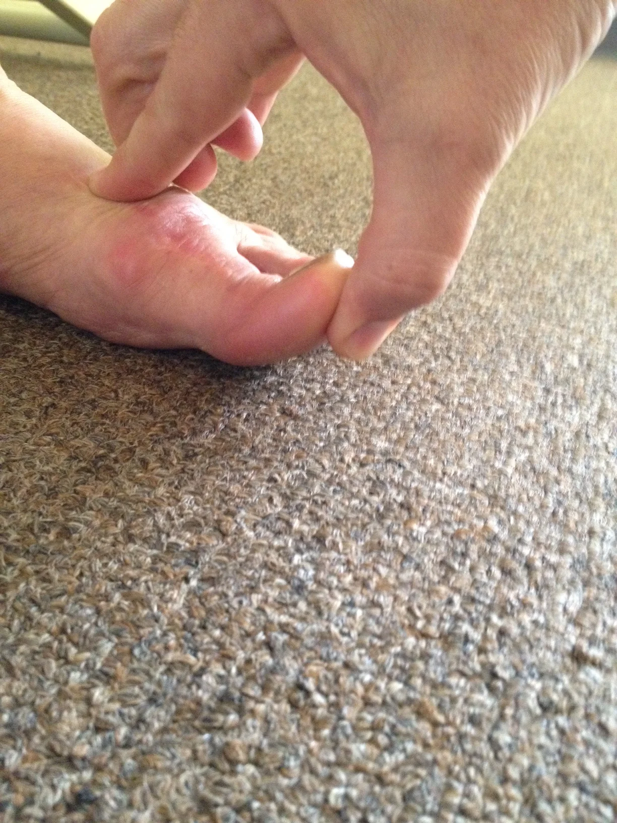
Here we have a very troubled foot. This foot has undergone numerous procedures, sadly. Today we will not talk about the hallux varus you see here, a virtual unicorn in practice  (and acquired in this case) nor do we want to discuss the phalangeal varus drift. We want to draw your attention to the obvious impairment of the 1st MTP (metatarsophalangeal joint) dorsiflexion range.  You can see the large dorsal crown of osteophytes, a dorsal buttress to any hallux dorsiflexion.  There is under 10 degrees of dorsiflexion here, not even enough worth mentioning.  We have said it many times before, if you lose a range at one joint usually that range has to be accommodated for proximal or distal to the impaired joint. This is a compensation pattern and you can see it here in the hallux joints themselves.

Here you can see that some of the dorsiflexion range has been acquired in the proximal phalangeal joint.  We like to call this “banana toe” when explaining it to patients, it is a highly technical term but you are welcome to borrow it. This occurred because the joint was constantly seeing the limitation of dorsiflexion of the 1st MTP joint and seeing, and accommodating to, the demands of the need for more dorsiflexion at toe off. 

But, here is the kicker. You have likely seen this video of ours on Youtube on how to acquire a foot tripod from using the toe extensors to raise the arch.  Video link here  and here.  Well, in his patient’s case today, they have a limitation of 1st MTP dorsiflexion, so the ability to maximally raise the arch is impaired. The Windlass mechanism is broken; “winding” of the plantar fascia around the !st MTP mechanism is not sufficiently present. Any limitations in toe extension (ie dorsiflexion) or ankle dorsiflexion will mean that :

1. compensations will need to occur

2. The Windlass mechanism is insufficient

3. gait is impaired at distal swing phase and toe off phases

4. the anterior compartment competence will drop (Skill, endurance, strength) and thus injury can be more easily brought to the table.

In this patient’s case, they came in complaining of burning at the top of the foot and stiffness in the anterior ankle mortise area.  This would only come on after a long brisk walk.  If the walk was brisk yet short, no problems. If the walk was long and slow, no problems.  They clearly had an endurance problem and an endurance challenge in the office showed an immediate failure in under 30 seconds (we will try to shoot a quick video so show our little assessment so be patient with us). The point here today is that if there is a joint limitation, there will be a limitation in skill, strength or endurance and very likely a combination of the 3. If you cannot get to a range, then any skill, endurance or strength beyond that limitation will be lost and require a compensation pattern to occur.  This patient’s arch cannot be restored via the methods we describe here on our blog and it cannot be restored by an orthotic. The orthotic will likely further change, likely in a negative manner, the already limited function of the 1st MPJ. In other words, if you raise the arch, you will shorten the plantar fascia and draw the 1st MET  head towards the heel (part of the function of the Windlass mechanism) and by doing this you will plantarflex the big toe … .  but weren’t we praying for an increase in dorsiflexion of the limitus big toe ? ……..yes, exactly !  So use your head  (and spank the orthotic when you see it used in this manner.  ”Bad orthotic, bad orthotic !”)

So think of all of this the next time you see a turf toe / hallux rigidus/ hallux limitus. Rattles your brain huh !?

This is not stuff for the feint of heart. You gotta know your biomechanics.

Shawn and Ivo … .the gait guys

Addendum for clarity:

a Facebook reader asked a question:

From your post: “if you raise the arch, you will shorten the plantar fascia and draw the 1st MET head towards the heel (part of the function of the Windlass mechanism) and by doing this you will plantarflex the big toe … . but weren’t we praying for an increase in dorsiflexion of the limitus big toe ? ” I always thought when the plantar fascia is shortened, it plantar flexes the 1st metatarsal (1st ray) and extends (dorsiflexes) the 1st MTP joint….

Our response:  

We should have been more clear, our apologies dear reader.  Here is what we should have said , ” The plantar fascia is non-contractile, so it does not shorten. We meant conceptually shorten. When in late stance phase, particularly at toe off when the heel has raised and forefoot loading is occurring, the Windlass mechanism around the 1st MET head (as the hallux is dorsiflexing) is drawing the foot into supination and thus the heel towards the forefoot (ie passive arch lift). This action is driving the 1st MET into plantarflexion in the NORMAL foot.  This will NORMALLy help with increasing hallux dorsiflexion. In this case above, there is a rigid 1st MTP  joint.  So this mechanism cannot occur at all. In this case the plantar fascia will over time retract to the only length it does experience. So, if an orthotic is used, it will press up into the fascia and also plantarflex the 1st MET, which will carry the rigid toe into plantar flexion with it, IN THIS CASE.”

Podcast 51: Bouncy Gait, Stem Cells & Plantar fasciitis,

A. Link to our server:

http://thegaitguys.libsyn.com/podcast-51-bouncy-gait-stem-cells-plantar-fasciitis

B. iTunes link:

https://itunes.apple.com/us/podcast/the-gait-guys-podcast/id559864138

C. Gait Guys online /download store (National Shoe Fit Certification and more !) :

http://store.payloadz.com/results/results.aspx?m=80204

D. other web based Gait Guys lectures:

www.onlinece.com   type in Dr. Waerlop or Dr. Allen,  ”Biomechanics”

________________________________________

* Today’s show notes:

Neuroscience:

1. Stem cells and plantar fasciitis.
2. Study finds axon regeneration after Schwann cell graft to injured spinal cord
http://www.medicalnewstoday.com/releases/270623.php
3.Future Tiny Robots Will Communicate Using Only Molecules
http://www.fastcoexist.com/3020657/futurist-forum/future-tiny-robots-will-communicate-using-only-molecules?partner=rss
Correction:
a. I was listening to pod 49, great job. I am sending a personal message to point out an error regarding body composition. Under water weighing is considered the 2 compartment “Gold Standard” however caliper testing is used more due to cost and ability to be used in the field. Loved the learning stuff I sent some stuff similar to what you were talking about to the school administration but it went no where.
Thanks guys, Mark
b. Mark wrote: “I can see widening base of support to increase stability when one is weak but to widen base of support when one has decreased traction may increase slipping depending on width of stance couldn’t it. If decreased traction is a issue wouldn’t a better statagy”
Blog reader:
Not very infrequently the foot tripod has been discussed. Especially the importance of the medial tripod (MT) has been of great benefit to me. Some kind of a peroneus paralysis was probably the cause of weak MT of the right foot. With a weak anterior muscle group or compartment. But what about the lateral tripod, ie the 5th distal end of the fifth metatarsal. Which muscles are most responsible for the foot stability here and what kind of exercises might be of therapeutic value? Thank you.

Disclaimer
Gait Guys online /download store (National Shoe Fit Certification and more !) :

http://store.payloadz.com/results/results.aspx?m=80204 

Dear Gait Guys,                                
I’ve had a problem for a while where my lower left leg whips across the midline of my body at the start of the swing phase. This happens immediately after my left foot leaves the ground and before my right foot makes contact. My left knee seems to be angled outward, and I think this is due to some sort of external rotation of the hip or femur during the the early part of my swing phase. I attached a picture to illustrate this problem in my gait. I recently came across a blog post you guys wrote (http://thegaitguys.tumblr.com/post/14262793786/gait-problem-the-solitary-externally-rotated) and under the possibilities as to why there isn’t enough internal rotation, the second option describes exactly what happens when I run. So my question is, what can be done to correct this improper gait pattern? Thank you very much for taking the time to read this.
Sincerely,
Matthew
Between a quarter and a third of everything on the web is copied from somewhere else


tumblr_m3twiqKxiS1qhko2so1_500.jpg
tumblr_m3twiqKxiS1qhko2so2_500.jpg
Plantar Fascitis?  
 
You’ve got plantar fascitis? We’ll try steroid injections. If that does not work, no problem, we’ll just cut it out…. 
 
Ah, yes…..Nothing like cutting one of the main stabilizing influences for the foot (via the windlass mechanism) to accomplish your goals. We sure are glad they used dead feet in this study!                 
And now, here is more evidence that those ligaments play a significant role (along, of course, with competent musculature) in stability of the foot.    

          The conclusion: “The data suggest that operations involving fasciotomy affect arch stability and should not be performed in patients with evidence of concomitant pes planus deformity, because of the likelihood of further deformation.”    
 
                                                                                                                                   The Gait Guys: Just the facts, so you can make more educated decisions..                                                              

                                                                                                                                  Foot Ankle Int. 1997 Jan;18(1):8-15.
Mechanical behavior of the foot and ankle after plantar fascia release in the unstable foot.

Kitaoka HB, Luo ZP, An KN.

Department of Orthopedics, Mayo Clinic, Rochester, Minnesota, USA.

Abstract

The change in position of the bones of the foot was studied in three dimensions after plantar fascia release in intact and destabilized feet. Fifteen fresh-frozen human foot specimens were used. Physiologic loads of 445 newtons were applied axially to simulate standing at ease, and the three-dimensional position of tarsal bones was determined with a magnetic tracking device. The positions were presented in the form of screw axis displacements, quantitating rotation, and axis of rotation orientation. After fasciotomy in the six intact feet, significant differences in rotation were observed at the talotibial and calcaneotalar levels. After fasciotomy in the four unstable feet with three supporting elements sectioned, significant differences in position were observed at the talotibial joint and a significant decrease in arch height was observed. After fasciotomy in the five unstable feet with five supporting elements sectioned, significant differences in rotation were observed at the talotibial joint (mean, 5.5 +/- 1.6 degrees; P = 0.001), calcaneotalar joint (mean, 6.1 +/- 2.1 degrees; P = 0.003), and metatarsotalar level (mean, 9.3 +/- 4.1 degrees; P = 0.007). The average decrease in arch height was 7.4 +/- 4.1 mm (P = 0.015). Displacement of all joints tested occurred after fasciotomy, with rotation about all three axes. These changes in displacement were more pronounced in unstable or destabilized feet. The data suggest that operations involving fasciotomy affect arch stability and should not be performed in patients with evidence of concomitant pes planus deformity, because of the likelihood of further deformation.

http://www.ncbi.nlm.nih.gov/pubmed/9013108