tumblr_no75l1nCgP1qhko2so1_r1_400.jpg
tumblr_no75l1nCgP1qhko2so2_r1_400.jpg

En Pointe, Demi Pointe, Posterior Impingement ?

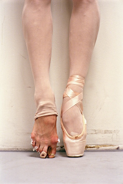
When we see pictures like this most of us are triggered to look at the toe and the challenges to the 1st MTP joint.  But what about all that compression and crowding in the back of the ankle ? Posterior compression is a reality in athletes who spend time at end range plantarflexion or pack much force and load through end range plantarflexion.

This is a photo example of what is referred to as “en pointe” which means “on the tip”.  “Demi pointe” means on the ball of the foot which is much safer for many areas of the foot, but this requires adequate 1st MTP (metatarsophalangeal joint range). We discussed this briefly this week on social media regarding hallux limitus and rigidus.

En Pointe is a terrible challenge. So if you are thinking of putting your darling children in ballet…… just beware of the facts and do some logical thinking on your own when it comes to allowing the “en pointe” axially loading of the entire body over a single joint, a type of loading that this joint was never, ever, designed to withstand. This joint is a great problem for a great many in their lives, why start playing with the risk factors so early ? Let them dance, into demi pointe, but pull them once they are being forced in to En Pointe, if you want our opinion on the matter.

En pointe or classical point ballet it typically done in point shoes or slippers which have a reinforced toe box that allows a more squared off stable surface to stand in pointe position.  It does not however allow a reduction in the axial loading that you see in this picture and it certainly does not help with proper angulation of the big toe, if anything the slipper will gently corral the toes together rendering abductor hallucis muscle function nearly obsolete.   The box will also not stop the valgus loading that typically occurs at the joint. Despite what the studies say, this is one we would watch carefully.  Now, there are studies out there that do not support hallux valgus and bunion formation in dancers, we admit that.  However, we are just asking you to use common sense.  If you see a bunion forming, if the toe is getting chronically swollen, if the toe is drifting off line then one must use common sense and assume that the load is exceeding joint integrity.  Prolonged and excessive loading of any joint cartilage is likely to create a risky environment to crack, fissure, wear down or damage the cartilage or the bony surface underneath (subchondral bone).  If you screw up this joint, toe off will be impaired and thus the windlass effect at the joint will be impaired thus leading to a multitude of other dysfunctional foot issues in the years to come.

Now, back to the “en pointe” position.  Did you try it yet ? Heed our warning ! Just trust us, this is bloody hard.  Since serious foot deformities can result from starting pointe too early, pre-professional students do not usually begin dancing en pointe until after the age of 10 or so , remember, the adolescent foot has not completed its bone ossification and the bone growth plates have not closed.  Thus, damage and deformity are to be expected if done at too young an age.  If you asked our opinion on this, we would say to wait until at least the mid-teenage years……. but by that point in the dance world a prodigy would miss her or his opportunity.  Thus, we see the problems from going “en pointe” too early in many. In the dance world, there are other qualifications for dancers before En Pointe is begun. Things like holding turnout, combining center combinations, secure and stable releve, 3rd position, 4th position, 4th croise and 5th position all of which are huge torsional demands on the hips to the feet. Do you want your child undergoing these deforming forces during early osseous development ? 

Achieving en pointe is a process.  There is a progression to get to it.  Every teacher has their own methods but it is not a “just get up on your toes” kind of thing.

Are you a dancer with posterior ankle pain, impingement or disability. The Os trigonum and protruding lateral talar process are two common and well-documented morphological variations associated with posterior ankle impingement in ballet dancers. 

Think this stuff through. If you are going to be treating these things, you have to know the anatomy, loading mechanics and you have to know your sport or art. Dr. Allen was a physician for the world famous Joffrey Ballet for a few years, he knows a thing or two about these issues dancer’s endure. And he still has a few nightmares from time to time over them. 

Dr. Shawn Allen

reference:

Clin Anat. 2010 Sep;23(6):613-21. doi: 10.1002/ca.20991.Pathoanatomy of posterior ankle impingement in ballet dancers. Russell JA,Kruse DW, Koutedakis Y, McEwan IM, Wyon M

A case of the non-resolving ankle sprain.  Things to think about when the ankle and foot just do not fully come around after a sprain.
Gait Guys,
A while back I had a severe ankle sprain while trail running.  As I stepped on a rock my toes pointed d…

A case of the non-resolving ankle sprain.  Things to think about when the ankle and foot just do not fully come around after a sprain.

Gait Guys,

A while back I had a severe ankle sprain while trail running.  As I stepped on a rock my toes pointed downward, my ankle was rolled in and I felt a pop. This was follow by a lot of swelling and bruising both on the inside and outside of my ankle.  Being experienced with ankle sprains, I jumped on the initial treatment immediately. The reduction in swelling and bruising lead me to believe that I was in for a 4-5 week recovery, then I would be back at what I love doing. I was proven wrong:  

1.       Initial treatment consisted of immobilization, icing, and a very high dose of Ibuprofen (3 days only). After a couple weeks of this I began stretching, massage and trying to get into some modified activities as the pain allowed me to. I was able to  do some hiking but running was too painful.

2.       After 6 weeks, I was still having pain in the posterior tibial tendon area as well as the deltoid ligament area. I tried running but, I was met with severe pain beginning in the middle of the gait cycle through  the push off. I saw a PA at this time and was told to give it more rest. For the next few weeks I wore a soft brace and spent most of my time in a chair.

3.       By week 9, there was no improvement. I could walk fine but, I had the same pain when I tried to run. I visited the PA again and was put in a walking cast and had an MRI. The MRI should a low grade deltoid and ATFL sprain as well as a bruised bone. I spent 2 weeks in the walking cast then returned to the soft cast for another week. During this time I did nothing besides give it rest.

4.       At week 11, I did not see a noticeable improvement. I still had a sharp pain in my posterior tibial tendon area and deltoid area during the middle of my gait (when trying to run). At this time, I had another visit with the PA. After looking at my MRI more closely, he saw fluid buildup behind my talus. He thinks that I had an impact injury to my Os Trigonum. He also noticed that I had very limited dorsiflexion.  He has advised me to stretch and give it a few more weeks. If it’s not going in a positive direction he recommended a cortisone shot.

As it stands today at week 12, in a dorsiflexion position, I have a sharp pain in what feels like my Achilles tendon and posterior tibial tendon area (the MRI shows these are intact). I also have a lot of tenderness in the deltoid area. Walking, I am almost pain free but as soon as I begin to run, the pain starts in the areas described above. This is the first injury I have ever had where I haven’t seen a steady improvement when recovering (maybe I am just getting old). The pain I am having now when trying to running is the same as it was at week 4. This really concerns me.

I guess my question is, where do I go from here? Do I keep doing what I am doing? Should I seek a second opinion?  Any help or guidance you could provide would be greatly appreciated.

On a side note, your blog has helped me to get though the last 12 run-less weeks without losing my mind or falling into a deep depression.  You guys do some great stuff.  Keep up the good work!

Best Regards,

MR

____________________

Dear MR:

Somehow we missed this email. Sorry about that.

Whenever things are not resolving with reasonable intervention one must think of two things: either the injury was severe or the diagnosis is incorrect.

Without seeing you we are unable to determine either. But here are our thoughts.

The Os Trigonum syndrome is a good thought. It seems to be in the correct area of your complaint. These “Os” bones can be embedded in tendon or soft tissue and they can be fixed to the posterior talus by either bone or a cartilagenous bridge. It is possible for this to be your problem if the inversion event was severe enough although it is not that common in this described mechanism.

One must also be suspect of osseous compression of the medial talus against the medial calcaneus, which will bring thoughts of a posterior subtalar facet fracture. We pulled up an article we read a few years ago on this issue (click here), the article is entitled, “Pseudo os trigonum sign: missed posteromedial talar facet fracture”. Obviously this needs to be considered in your case since there are similar components in area and symptom of your complaints. Posteromedial talar facet fracture (PMTFF) is a rare injury, sparsely reported in the literature and it must be chased as a diagnosis of suspicion when all other clinical presentations have not panned out. Damage to the sustentaculum tali must also be assessed, as this too can be fractured.  Osteochondral defects are also always on the list in violent inversion events; they are classically seen anteromedially and posteriolaterally at the ankle mortise joint.

Something else that is often missed in ankle inversion sprains is avulsion or rupture of the extensor digitorum brevis on the lateral foot. As the rearfoot inverts and forefoot plantarflexes the EDB is tensioned to the point of tearing. Although you seem to have no symptoms in this area it can never be overlooked. These are easy to discern from the lateral ligamentous structure damage because the areas are clearly separate from eachother.  Look for tenderness down into the top of the metatarsals into the forefoot. Also test for weakness and pain of toe extension.

So, lots to consider here in this case. When things to not resolve you have to start looking for less common problems and damage.  We would love to hear how you are doing MR. Drop us a line.

Shawn and Ivo……. also geeks of orthopedics.  We paid the piper long ago.